Products
Research Areas
COVID-19
Resources
Login
Quick Order
Cart Cart lightblue
Login
Registration enables users to use special features of this website, such as past
order histories, retained contact details for faster checkout, review submissions, and special promotions.


Fields marked with a * are required.

Login
Quick Order
Contact Us

Location


Corporate Headquarters

Vector Laboratories, Inc.
6737 Mowry Ave
Newark, CA 94560
United States

Telephone Numbers


Customer Service: (800) 227-6666 / (650) 697-3600


Contact Us


Additional Contact Details

Login
Registration enables users to use special features of this website, such as past
order histories, retained contact details for faster checkout, review submissions, and special promotions.


Fields marked with a * are required.

Login
Quick Order

PDPK1 / PDK1

3-phosphoinositide dependent protein kinase-1

Serine/threonine kinase which acts as a master kinase, phosphorylating and activating a subgroup of the AGC family of protein kinases. Its targets include: protein kinase B (PKB/AKT1, PKB/AKT2, PKB/AKT3), p70 ribosomal protein S6 kinase (RPS6KB1), p90 ribosomal protein S6 kinase (RPS6KA1, RPS6KA2 and RPS6KA3), cyclic AMP-dependent protein kinase (PRKACA), protein kinase C (PRKCD and PRKCZ), serum and glucocorticoid-inducible kinase (SGK1, SGK2 and SGK3), p21-activated kinase-1 (PAK1), protein kinase PKN (PKN1 and PKN2). Plays a central role in the transduction of signals from insulin by providing the activating phosphorylation to PKB/AKT1, thus propagating the signal to downstream targets controlling cell proliferation and survival, as well as glucose and amino acid uptake and storage. Negatively regulates the TGF-beta-induced signaling by: modulating the association of SMAD3 and SMAD7 with TGF-beta receptor, phosphorylating SMAD2, SMAD3, SMAD4 and SMAD7, preventing the nuclear translocation of SMAD3 and SMAD4 and the translocation of SMAD7 from the nucleus to the cytoplasm in response to TGF-beta. Activates PPARG transcriptional activity and promotes adipocyte differentiation. Activates the NF-kappa-B pathway via phosphorylation of IKKB. The tyrosine phosphorylated form is crucial for the regulation of focal adhesions by angiotensin II. Controls proliferation, survival, and growth of developing pancreatic cells. Participates in the regulation of Ca2+ entry and Ca2+-activated K+ channels of mast cells. Essential for the motility of vascular endothelial cells (ECs) and is involved in the regulation of their chemotaxis. Plays a critical role in cardiac homeostasis by serving as a dual effector for cell survival and beta-adrenergic response. Plays an important role during thymocyte development by regulating the expression of key nutrient receptors on the surface of pre-T cells and mediating Notch-induced cell growth and proliferative responses. Provides negative feedback inhibition to toll-like receptor-mediated NF-kappa-B activation in macrophages. Isoform 3 is catalytically inactive.

Gene Name: 3-phosphoinositide dependent protein kinase-1
Family/Subfamily: Protein Kinase , RAC/AKT
Synonyms: PDPK1, HPDK1, PkB kinase like gene 1, PkB-like 1, Pdk-1, PkB kinase, PRO0461
Target Sequences: NM_002613 NP_002604.1 O15530

Publications (1)

1
Nox4 NAD(P)H oxidase mediates Src-dependent tyrosine phosphorylation of PDK-1 in response to angiotensin II: role in mesangial cell hypertrophy and fibronectin expression. Block K, Eid A, Griendling KK, Lee DY, Wittrant Y, Gorin Y. The Journal of biological chemistry. 2008 283:24061-76. (WB) [PubMed:18559349] [PMC:PMC2527116]

☰ Filters
Antibodies (64)
Proteins (10)
ELISA Kits (4)
Cell-Based (1)
Cell-Based Phosphorylation (1)
Over-Expression Lysate (4)
Primary (64)
Recombinant (6)
Sandwich (2)
PDPK1 / PDK1 (78)
Human (64)
Mouse (34)
Rat (32)
Bat (3)
Bovine (5)
Chimpanzee (3)
Dog (3)
Gibbon (3)
Guinea pig (4)
Monkey (5)
Pig (5)
Rabbit (2)
Sheep (2)
Zebrafish (3)
IHC (31)
IHC-P (14)
WB (50)
Flo (2)
DB (7)
ELISA (25)
ICC (1)
IF (6)
IP (2)
Peptide-ELISA (8)
rabbit (58)
mouse (6)
Product Group
IHCPlus (1)
IgG (40)
IgG1 (2)
IgG1,k (1)
IgG2a (1)
IgG2a,k (1)
IgM,k (1)
monoclonal mc (6)
polyclonal pc (58)
2E2 (1)
3H3D9 (1)
7E4G11 (1)
96-Well Microplate (2)
96-Well Strip Plate (2)
APC (1)
APC Conjugated (2)
Azide-free (1)
Biotin Conjugated (3)
Cy3 Conjugated (1)
Cy7 Conjugated (1)
FITC Conjugated (3)
HRP Conjugated (3)
PE Conjugated (2)
Unconjugated (49)
pSer241 (10)
aa83-338 (8)
pSer396 (7)
aa201-250 (4)
1-80 aa (2)
180-260 aa (2)
C-Terminus (2)
aa277-556 (2)
pTyr9 (2)
Internal (1)
Lys235 (1)
aa1-50 (1)
aa2-51 (1)
aa210-259 (1)
aa29-436 (1)
aa301-312 (1)
aa506-556 (1)
aa524-556 (1)
p241 (1)
pSer/Thr (1)
No (78)
Adherent Cell Cultures (2)
Cell Lysates (1)
Plasma (1)
Serum (1)
Tissue Homogenates (2)
GST (1)
GST, N-terminus (2)
His (1)
His-T7 (1)
Myc-DDK (Flag) (5)
Human (8)
293T Cells (2)
Baculovirus (1)
E. coli (1)
HEK 293 Cells (3)
Wheat Germ Extract (3)
Purified (2)
Low endotoxin level (1)
Select
PDPK1 / PDK1 Rabbit anti-Human Polyclonal (aa506-556) Antibody
Mouse, Human
IP, WB
Unconjugated
10 µl/$295; 100 µl/$514
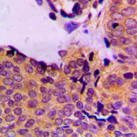
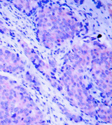
PDPK1 / PDK1 Antibody - Immunohistochemical analysis of PDPK1 (pS241) staining in human breast cancer formalin fixed paraffin embedded tissue section. The section was pre-treated using heat mediated antigen retrieval with sodium citrate buffer (pH 6.0). The section was then incubated with the antibody at room temperature and detected using an HRP conjugated compact polymer system. DAB was used as the chromogen. The section was then counterstained with hematoxylin and mounted with DPX.
Select
PDPK1 / PDK1 Rabbit anti-Human Polyclonal (pSer241) Antibody
Rabbit, Mouse, Sheep, Bovine, Rat, Pig, Human
IHC, IHC-P, WB
Unconjugated
100 µl/$315
Select
PDPK1 / PDK1 Rabbit anti-Human Polyclonal (pSer241) Antibody
Mouse, Rat, Human
IF, IHC, WB
Unconjugated
50 µg/$489
Select
PDPK1 / PDK1 Mouse anti-Human Monoclonal (pSer/Thr) Antibody
ELISA, IP, WB
Unconjugated
100 µl/$689
Select
PDPK1 / PDK1 Mouse anti-Human Monoclonal (7E4G11) Antibody
Human
ELISA, Flo, IF, IHC, WB
Unconjugated
100 µl/$459
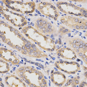
PDPK1 / PDK1 Antibody - Immunohistochemistry of paraffin-embedded human kidney using PDPK1 Antibodyat dilution of 1:200 (40x lens).
Select
PDPK1 / PDK1 Rabbit anti-Human Polyclonal Antibody
Mouse, Human
IF, IHC, WB
Unconjugated
20 µl/$287; 100 µl/$431
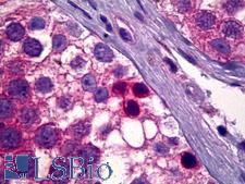
PDPK1 / PDK1 Antibody - Anti-PDK1 antibody IHC of human testis, spermatogonia. Immunohistochemistry of formalin-fixed, paraffin-embedded tissue after heat-induced antigen retrieval. Antibody concentration 5 ug/ml.
Select
PDPK1 / PDK1 Rabbit anti-Human Polyclonal Antibody
Human
ELISA, IHC, IHC-P, WB
Unconjugated
50 µl/$460
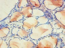
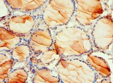
PDPK1 / PDK1 Antibody - Immunohistochemistry of paraffin-embedded human thyroid using antibody at 1:100 dilution.
Select
PDPK1 / PDK1 Rabbit anti-Human Polyclonal (aa277-556) Antibody
Human
ELISA, IHC, IHC-P, WB
Unconjugated
50 µl/$294; 100 µl/$360
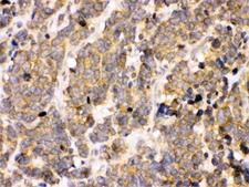
PDPK1 / PDK1 Antibody - Immunohistochemical analysis of paraffin-embedded human breast carcinoma tissue.
Select
PDPK1 / PDK1 Rabbit anti-Human Polyclonal (pSer241) Antibody
IF, IHC, WB
Unconjugated
20 µl/$287; 100 µl/$431
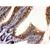
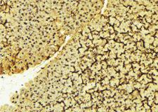
PDPK1 / PDK1 Antibody - PDPK1 antibody IHC-paraffin. IHC(P): Mouse Intestine Tissue.
Select
PDPK1 / PDK1 Rabbit anti-Human Polyclonal (aa524-556) Antibody
Mouse, Guinea pig, Rat, Human
IHC, IHC-P, WB
Unconjugated
10 µg/$318; 100 µg/$470
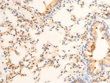
PDPK1 / PDK1 Antibody - 1:100 staining mouse lung tissue by IHC-P. The tissue was formaldehyde fixed and a heat mediated antigen retrieval step in citrate buffer was performed. The tissue was then blocked and incubated with the antibody for 1.5 hours at 22°C. An HRP conjugated goat anti-rabbit antibody was used as the secondary.
Select
PDPK1 / PDK1 Rabbit anti-Human Polyclonal (pSer241) Antibody
Mouse, Rat, Human
IHC, Peptide-ELISA, WB
Unconjugated
100 µl/$379; 200 µl/$421
PDPK1 / PDK1 Antibody - Immunohistochemistry of paraffin-embedded human thyroid using antibody at 1:100 dilution.
Select
PDPK1 / PDK1 Rabbit anti-Human Polyclonal (aa277-556) Antibody
Human
ELISA, IHC, IHC-P
Unconjugated
50 µl/$294; 100 µl/$360
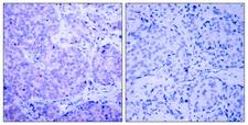
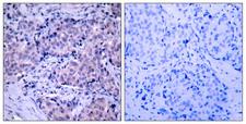
PDPK1 / PDK1 Antibody - Immunohistochemistry analysis of paraffin-embedded human breast carcinoma, using PDK1 (Phospho-Ser241) Antibody. The picture on the right is blocked with the phospho peptide.
Select
PDPK1 / PDK1 Rabbit anti-Human Polyclonal (pSer241) Antibody
Mouse, Rat, Human
IHC, IHC-P, Peptide-ELISA, WB
Unconjugated
50 µl/$334; 100 µl/$397
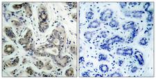
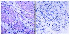
PDPK1 / PDK1 Antibody - Immunohistochemistry analysis of paraffin-embedded human breast carcinoma tissue, using PDK1 Antibody. The picture on the right is blocked with the synthesized peptide.
Select
PDPK1 / PDK1 Rabbit anti-Human Polyclonal (aa210-259) Antibody
Mouse, Rat, Human
IHC, IHC-P, Peptide-ELISA, WB
Unconjugated
50 µl/$334; 100 µl/$397
PDPK1 / PDK1 Antibody
Select
PDPK1 / PDK1 Rabbit anti-Human Polyclonal Antibody
Mouse, Rat, Human
IHC, IHC-P, WB
Unconjugated
100 µg/$575
Select
PDPK1 / PDK1 Rabbit anti-Human Polyclonal (1-80 aa) Antibody
Mouse, Rat, Human, Monkey
ELISA, IHC, WB
Unconjugated
50 µg/$295; 100 µg/$335; 200 µg/$394
Select
PDPK1 / PDK1 Rabbit anti-Human Polyclonal (180-260 aa) Antibody
Mouse, Rat, Human
ELISA, IHC, WB
Unconjugated
50 µg/$295; 100 µg/$335; 200 µg/$394
PDPK1 / PDK1 Antibody - 1:100 staining mouse liver tissue by IHC-P. The sample was formaldehyde fixed and a heat mediated antigen retrieval step in citrate buffer was performed. The sample was then blocked and incubated with the antibody for 1.5 hours at 22°C. An HRP conjugated goat anti-rabbit antibody was used as the secondary.
Select
PDPK1 / PDK1 Rabbit anti-Human Polyclonal Antibody
Mouse, Rat, Human
IHC, Peptide-ELISA, WB
Unconjugated
100 µl/$379; 200 µl/$421
PDPK1 / PDK1 Antibody - Western blot of affinity purified anti-PKD-1 antibody shows detection of myc-tagged human PDK-1 at 60kD in ~10 ug of a virus infected Sf9 cell lysate (arrow). The nitrocellulose membrane was probed overnight at 4 degrees C with the primary antibody diluted 1:750 in 1% non-fat dry milk. HRP conjugated Goat-anti-Rabbit IgG and chemiluminescent detection is recommended. Other detection systems will yield similar results.
Select
PDPK1 / PDK1 Rabbit anti-Human Polyclonal (C-Terminus) Antibody
Human
ELISA, IHC, WB
Unconjugated
100 µg/$567
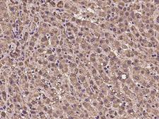
PDPK1 / PDK1 Antibody - Immunochemical staining of human PDPK1 in human liver with rabbit polyclonal antibody at 1:100 dilution, formalin-fixed paraffin embedded sections.
Select
PDPK1 / PDK1 Rabbit anti-Human Polyclonal Antibody
Human
ICC, IF, IHC-P
Unconjugated
100 µl/$412
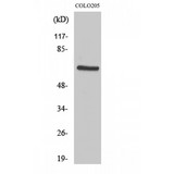
PDPK1 / PDK1 Antibody - Western blot of PDK1 antibody
Select
PDPK1 / PDK1 Rabbit anti-Human Polyclonal (180-260 aa) Antibody
Mouse, Rat, Human
ELISA, IHC, WB
Unconjugated
50 µg/$295; 100 µg/$335; 200 µg/$394
PDPK1 / PDK1 Antibody - Peptide - + Immunohistochemical analysis of paraffin- embedded human breast carcinoma tissue using PDK1 (Ab-241) antibody.
Select
PDPK1 / PDK1 Rabbit anti-Human Polyclonal (p241) Antibody
Mouse, Rat, Human
ELISA, IHC, WB
Unconjugated
100 µg/$357
PDPK1 / PDK1 Antibody - P-Peptide - + Immunohistochemical analysis of paraffin- embedded human breast carcinoma tissue using PDK1 (phospho-Ser241) antibody.
Select
PDPK1 / PDK1 Rabbit anti-Human Polyclonal (pSer241) Antibody
Mouse, Rat, Human
ELISA, IHC, WB
Unconjugated
100 µg/$357
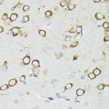
PDPK1 / PDK1 Antibody - Immunohistochemistry of paraffin-embedded Rat brain using PDPK1 Polyclonal Antibody at dilution of 1:200 (40x lens).
Select
PDPK1 / PDK1 Rabbit anti-Human Polyclonal Antibody
Mouse, Rat, Human
IF, IHC
Unconjugated
60 µl/$343; 120 µl/$427; 200 µl/$559
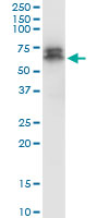
PDPK1 / PDK1 Antibody - PDPK1 monoclonal antibody (M05), clone 2E2. Western Blot analysis of PDPK1 expression in rat brain.
Select
PDPK1 / PDK1 Mouse anti-Human Monoclonal (2E2) Antibody
Rat, Human
ELISA, WB
Unconjugated
100 µg/$423
Viewing 1-25 of 78 product results


If you do not find the reagent or information you require, please contact Customer.Support@LSBio.com to inquire about additional products in development.

PLEASE NOTE

For RESEARCH USE ONLY. Intended for use by laboratory professionals. Not intended for human diagnostic or therapeutic purposes.

The data on this page has been compiled from LifeSpan internal sources, the National Center for Biotechnology Information (NCBI), and The Universal Protein Resource (UniProt).