Login
Registration enables users to use special features of this website, such as past
order histories, retained contact details for faster checkout, review submissions, and special promotions.
order histories, retained contact details for faster checkout, review submissions, and special promotions.
Forgot password?
Registration enables users to use special features of this website, such as past
order histories, retained contact details for faster checkout, review submissions, and special promotions.
order histories, retained contact details for faster checkout, review submissions, and special promotions.
Quick Order
Products
Antibodies
ELISA and Assay Kits
Research Areas
Infectious Disease
Resources
Purchasing
Reference Material
Contact Us
Location
Corporate Headquarters
Vector Laboratories, Inc.
6737 Mowry Ave
Newark, CA 94560
United States
Telephone Numbers
Customer Service: (800) 227-6666 / (650) 697-3600
Contact Us
Additional Contact Details
Login
Registration enables users to use special features of this website, such as past
order histories, retained contact details for faster checkout, review submissions, and special promotions.
order histories, retained contact details for faster checkout, review submissions, and special promotions.
Forgot password?
Registration enables users to use special features of this website, such as past
order histories, retained contact details for faster checkout, review submissions, and special promotions.
order histories, retained contact details for faster checkout, review submissions, and special promotions.
Quick Order
LSBio Phospho-Specific ELISA Kits
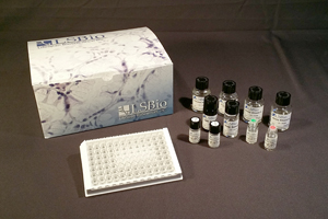
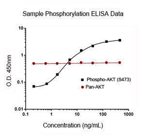
LifeSpan offers a number of different types of ELISA kit that are specific for the detection of phosphorylated target proteins.
Phospho-specific kits are available for standard ELISA detection, in vitro detection in adherent cells, and detecting transcription
factor activity in nuclear extracts and cell lysates. Each kit is ready-to-use with all the necessary reagents and a clear, concise
protocol that will step you through the process, from sample preparation to analysis of the results.
Phospho-Specific Sandwich ELISA Kit Protocol
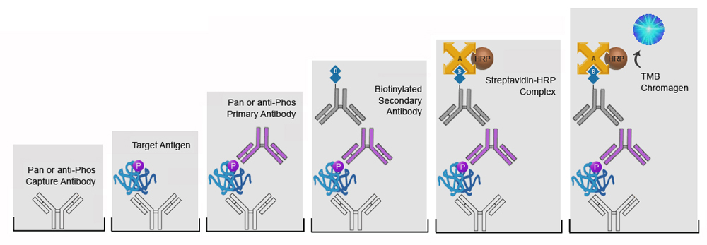
| 1) The supplied 96-well microtiter plate is pre-coated either pan- or phospho-specific capture antibody. |
| 2) Control standards or test samples are added to the well and the target antigen is captured by the antibodies. |
| 3) Phospho-specific or pan-specific primary antibody is added and binds to the target antigen. |
| 4) The wells are washed and a biotinylated secondary antibody is added and binds to the primary antibody. |
| 5) The wells are washed and a solution containing HRP-conjugated Avidin is added. The Avidin binds to the biotinylated secondary antibody. |
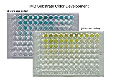
| 6) The wells are washed and a solution containing TMB is added. The TMB reacts with the HRP changing from colorless to a blue color. An acid stop solution is then added, the reaction stops, and the blue color changes to yellow. |
Phospho-Specific DNA-Binding ELISA Kit Protocol

| 1) The 96-well microtiter plate comes pre-bound with specific double-stranded (dsDNA) oligonucleotides. |
| 2) Following a blocking step samples are added to each well and phosphorylated transcription factor binds to the oligonucleotides. |
| 3) The wells are washed and a phospho-specific primary antibody is added which binds activated transcription factor. |
| 4) The wells are washed and a HRP-conjugated secondary antibody is added which binds to the primary antibody. |
| 5) The wells are washed and a solution containing TMB is added. The TMB reacts with the HRP changing from colorless to a blue color. An acid stop solution is then added, the reaction stops, and the blue color changes to yellow. |
Phospho-Specific Cell-Based ELISA Kit Protocol

| 1) Cell monolayers are cultured in the 96-well microtiter plate. Each well can then be treated with stimulants, inhibitors, etc. to induce expression of the phosphorylated target. The cells are then fixed and blocked. |
| 2) Phospho-specific or pan-specific primary antibody is added and binds to the target antigen on the cells surface. |
| 3) The wells are washed and a HRP-conjugated secondary antibody is added which binds to the primary antibody. |
| 4) The wells are washed and a solution containing TMB is added. The TMB reacts with the HRP changing from colorless to a blue color. An acid stop solution is then added, the reaction stops, and the blue color changes to yellow. |
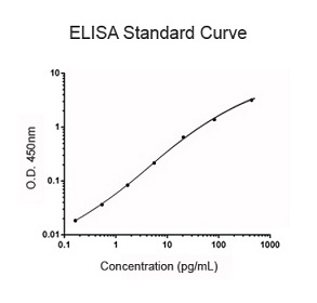
The plate can then be read in a spectrophotometer at a wavelength of 450 nm.
