Products
Research Areas
COVID-19
Resources
Login
Quick Order
Cart Cart lightblue
Login
Registration enables users to use special features of this website, such as past
order histories, retained contact details for faster checkout, review submissions, and special promotions.


Fields marked with a * are required.

Login
Quick Order
Contact Us

Location


Corporate Headquarters

Vector Laboratories, Inc.
6737 Mowry Ave
Newark, CA 94560
United States

Telephone Numbers


Customer Service: (800) 227-6666 / (650) 697-3600


Contact Us


Additional Contact Details

Login
Registration enables users to use special features of this website, such as past
order histories, retained contact details for faster checkout, review submissions, and special promotions.


Fields marked with a * are required.

Login
Quick Order

GFP

Green Fluorescent Protein (GFP)

Green Fluorescent Protein (GFP) exhibits bright green fluorescence when exposed to blue light. Although many other marine organisms have similar green fluorescent proteins, GFP traditionally refers to the protein first isolated from the jellyfish Aequorea victoria. GFP from A. victoria has a major excitation peak at a wavelength of 395 nm and a minor one at 475 nm. Its emission peak is at 509 nm, which is in the lower green portion of the visible spectrum. GFP from the sea pansy (Renilla reniformis) has a single major excitation peak at 498 nm. In cell and molecular biology, the GFP gene is frequently used as a reporter of expression. In modified forms it has been used to make biosensors, and many animals have been created that express GFP as a proof-of-concept that a gene can be expressed throughout a given organism. The GFP gene can be introduced into organisms and maintained in their genome through breeding, injection with a viral vector, or cell transformation.

Gene Name: Green Fluorescent Protein (GFP)
Synonyms: GFP
Target Sequences: M62653 P42212 P42212

Publications (32)

1
Direct interaction between the actin-binding protein filamin-A and the inwardly rectifying potassium channel, Kir2.1. Sampson LJ, Leyland ML, Dart C. The Journal of biological chemistry. 2003 278:41988-97. [PubMed:12923176]
2
The Vif protein of HIV triggers degradation of the human antiretroviral DNA deaminase APOBEC3G. Conticello SG, Harris RS, Neuberger MS. Current biology : CB. 2003 13:2009-13. [PubMed:14614829]
3
Molecular cloning, characterization and tissue expression of porcine Toll-like receptor 4. Alvarez B, Revilla C, Chamorro S, Lpez-Fraga M, Alonso F, Domnguez J, Ezquerra A. Developmental and comparative immunology. 2006 30:345-55. [PubMed:16139355]
4
Type II epithelial cells are critical target for hyperoxia-mediated impairment of postnatal lung development. Yee M, Vitiello PF, Roper JM, Staversky RJ, Wright TW, McGrath-Morrow SA, Maniscalco WM, Finkelstein JN, O'Reilly MA. American journal of physiology. Lung cellular and molecular physiology. 2006 291:L1101-11. [PubMed:16861382]
5
Pseudomonas aeruginosa type III-secreted toxin ExoT inhibits host-cell division by targeting cytokinesis at multiple steps. Shafikhani SH, Engel J. Proceedings of the National Academy of Sciences of the United States of America. 2006 103:15605-10. [PubMed:17030800] [PMC:PMC1622869]
6
ORF73-null murine gammaherpesvirus 68 reveals roles for mLANA and p53 in virus replication. Forrest JC, Paden CR, Allen RD, Collins J, Speck SH. Journal of virology. 2007 81:11957-71. [PubMed:17699571] [PMC:PMC2168792] Related Antibodies: LS-C154208.
7
Identification of a novel mitotic phosphorylation motif associated with protein localization to the mitotic apparatus. Yang F, Camp DG, Gritsenko MA, Luo Q, Kelly RT, Clauss TR, Brinkley WR, Smith RD, Stenoien DL. Journal of cell science. 2007 120:4060-70. [PubMed:17971412] Related Antibodies: LS-C154208.
8
B-cell receptor activation inhibits AID expression through calmodulin inhibition of E-proteins. Hauser J, Sveshnikova N, Wallenius A, Baradaran S, Saarikettu J, Grundstrm T. Proceedings of the National Academy of Sciences of the United States of America. 2008 105:1267-72. [PubMed:18203819] [PMC:PMC2234127]
9
Aire controls the differentiation program of thymic epithelial cells in the medulla for the establishment of self-tolerance. Yano M, Kuroda N, Han H, Meguro-Horike M, Nishikawa Y, Kiyonari H, Maemura K, Yanagawa Y, Obata K, Takahashi S, Ikawa T, Satoh R, Kawamoto H, Mouri Y, Matsumoto M. The Journal of experimental medicine. 2008 205:2827-38. [PubMed:19015306] [PMC:PMC2585853]
10
Type A GABA-receptor-dependent synaptic transmission sculpts dendritic arbor structure in Xenopus tadpoles in vivo. Shen W, Da Silva JS, He H, Cline HT. The Journal of neuroscience : the official journal of the Society for Neuroscience. 2009 29:5032-43. [PubMed:19369572] [PMC:PMC2706946] Related Antibodies: LS-C154208.
11
Stabilizers of the Max homodimer identified in virtual ligand screening inhibit Myc function. Jiang H, Bower KE, Beuscher AE, Zhou B, Bobkov AA, Olson AJ, Vogt PK. Molecular pharmacology. 2009 76:491-502. [PubMed:19498040] [PMC:PMC2730382]
12
ProMAT calibrator: A tool for reducing experimental bias in antibody microarrays. Zangar RC, Daly DS, White AM, Servoss SL, Tan RM, Collett JR. Journal of proteome research. 2009 8:3937-43. [PubMed:19618941] Related Antibodies: LS-C154288.
13
Lef-1 isoforms regulate different target genes and reduce cellular adhesion. Jesse S, Koenig A, Ellenrieder V, Menke A. International journal of cancer. Journal international du cancer. 2010 126:1109-20. [PubMed:19653274] Related Antibodies: LS-C154208.
14
Splice variants of the dual specificity tyrosine phosphorylation-regulated kinase 4 (DYRK4) differ in their subcellular localization and catalytic activity. Papadopoulos C, Arato K, Lilienthal E, Zerweck J, Schutkowski M, Chatain N, Mller-Newen G, Becker W, de la Luna S. The Journal of biological chemistry. 2011 286:5494-505. [PubMed:21127067] [PMC:PMC3037663] Related Antibodies: LS-C154219.
15
The syntaxin 4 N terminus regulates its basolateral targeting by munc18c-dependent and -independent mechanisms. Torres J, Funk HM, Zegers MM, ter Beest MB. The Journal of biological chemistry. 2011 286:10834-46. [PubMed:21278252] [PMC:PMC3060534] Related Antibodies: LS-C154219.
16
Vaccinia extracellular virions enter cells by macropinocytosis and acid-activated membrane rupture. Schmidt FI, Bleck CK, Helenius A, Mercer J. The EMBO journal. 2011 30:3647-61. [PubMed:21792173] [PMC:PMC3181475] Related Antibodies: LS-C154219.
17
The role of PSD-95 and cypin in morphological changes in dendrites following sublethal NMDA exposure. Tseng CY, Firestein BL. The Journal of neuroscience : the official journal of the Society for Neuroscience. 2011 31:15468-80. [PubMed:22031893] Related Antibodies: LS-C154312.
18
Therapeutic gene delivery and transfection in human pancreatic cancer cells using epidermal growth factor receptor-targeted gelatin nanoparticles. Xu J, Amiji M. Journal of visualized experiments : JoVE. 2012 e3612. [PubMed:22231028] [PMC:PMC3400379]
19
Multi-compartmental nanoparticles-in-emulsion formulation for macrophage-specific anti-inflammatory gene delivery. Attarwala H, Amiji M. Pharmaceutical research. 2012 29:1637-49. (ELISA; Mouse) [PubMed:22281760]
20
Retinoblastoma tumor-suppressor protein phosphorylation and inactivation depend on direct interaction with Pin1. Rizzolio F, Lucchetti C, Caligiuri I, Marchesi I, Caputo M, Klein-Szanto AJ, Bagella L, Castronovo M, Giordano A. Cell death and differentiation. 2012 19:1152-61. [PubMed:22322860] [PMC:PMC3374078] Related Antibodies: LS-C154219.
21
Tuftsin-modified alginate nanoparticles as a noncondensing macrophage-targeted DNA delivery system. Jain S, Amiji M. Biomacromolecules. 2012 13:1074-85. (ELISA; Mouse) [PubMed:22385328]
22
High-throughput, multiplexed analysis of 3-nitrotyrosine in individual proteins. Jin H, Zangar RC. Current protocols in toxicology / editorial board, Mahin D. Maines (editor-in-chief) ... [et al.]. 2012 Unit 17.15. [PubMed:22511115] Related Antibodies: LS-C154219.
23
CD133+ melanoma subpopulations contribute to perivascular niche morphogenesis and tumorigenicity through vasculogenic mimicry. Lai CY, Schwartz BE, Hsu MY. Cancer research. 2012 72:5111-8. (ICC, IHC; Human) [PubMed:22865455] [PMC:PMC3463654]
24
Regulation of postnatal forebrain amoeboid microglial cell proliferation and development by the transcription factor Runx1. Zusso M, Methot L, Lo R, Greenhalgh AD, David S, Stifani S. The Journal of neuroscience : the official journal of the Society for Neuroscience. 2012 32:11285-98. (ICC; Mouse) [PubMed:22895712]
25
Identification of the Plasmodium berghei resistance locus 9 linked to survival on chromosome 9. Bopp SE, Rodrigo E, González-Páez GE, Frazer M, Barnes SW, Valim C, Watson J, Walker JR, Schmedt C, Winzeler EA. Malaria journal. 2013 12:316. (IHC; Mouse) [Full Text Article] [PubMed:24025732] [PMC:PMC3848760] Related Antibodies: LS-C67081.
26
Dystroglycan controls dendritic morphogenesis of hippocampal neurons in vitro. Bijata M, Wlodarczyk J, Figiel I. Frontiers in cellular neuroscience. 2015 9:199. (IHC; Aequorea victoria) [Full Text Article] [PubMed:26074769] [PMC:PMC4443029] Related Antibodies: LS-C50850.
27
Anks1a regulates COPII-mediated anterograde transport of receptor tyrosine kinases critical for tumorigenesis. Lee H, Noh H, Mun J, Gu C, Sever S, Park S. Nature communications. 2016 7:12799. (IF; Human) [Full Text Article] [PubMed:27619642] [PMC:PMC5027278]
28
Hepatocellular Carcinoma Cell-Secreted Exosomal MicroRNA-210 Promotes Angiogenesis In Vitro and In Vivo. Lin XJ, Fang JH, Yang XJ, Zhang C, Yuan Y, Zheng L, Zhuang SM. Molecular therapy. Nucleic acids. 2018 June;11:243-252. (Aequorea victoria) [Full Text Article] [PubMed:29858059] [PMC:PMC5992447] Related Antibodies: LS-C154219.
29
The nicotinic a6 subunit gene determines variability in chronic pain sensitivity via cross-inhibition of P2X2/3 receptors. Wieskopf JS, Mathur J, Limapichat W, Post MR, Al-Qazzaz M, Sorge RE, Martin LJ, Zaykin DV, Smith SB, Freitas K, Austin JS, Dai F, Zhang J, Marcovitz J, Tuttle AH, Slepian PM, Clarke S, Drenan RM, Janes J, Al Sharari S, Segall SK, Aasvang EK, Lai W, Bittner R, Richards CI, Slade GD, Kehlet H, Walker J, Maskos U, Changeux JP, Devor M, Maixner W, Diatchenko L, Belfer I, Dougherty DA, Su AI, Lummis SC, Imad Damaj M, Lester HA, Patapoutian A, Mogil JS. Science translational medicine. 2015 May;7:287. (Aequorea victoria) [Full Text Article] [PubMed:25972004] [PMC:PMC5018401]
30
Fibrotic microenvironment promotes the metastatic seeding of tumor cells via activating the fibronectin 1/secreted phosphoprotein 1-integrin signaling. Zhang C, Wu M, Zhang L, Shang LR, Fang JH, Zhuang SM. Oncotarget. 2016 July;7:45702-45714. (Aequorea victoria) [Full Text Article] [PubMed:27329720] [PMC:PMC5216754] Related Antibodies: LS-C154219.
31
Detection of membrane protein-protein interaction in planta based on dual-intein-coupled tripartite split-GFP association. Tzu-Yin Liu, Wen-Chun Chou, Wei-Yuan Chen, Ching-Yi Chu, Chen-Yi Dai, Pei-Yu Wu. The Plant journal : for cell and molecular biology. 2018 May;94:426-438. [Full Text Article] [PubMed:29451720]
32
DHHC7-mediated palmitoylation of the accessory protein barttin critically regulates the functions of ClC-K chloride channels. Nataliya Gorinski, Daniel Wojciechowski, Daria Guseva, Dalia Abdel Galil, Franziska E Mueller, Alexander Wirth, Stefan Thiemann, Andre Zeug, Silke Schmidt, Monika Zareba-Koziol, Jakub Wlodarczyk, Boris V Skryabin, Silke Glage, Martin Fischer, Samer Al-Samir, Nicole Kerkenberg, Christa Hohoff, Weiqi Zhang, Volker Endeward, Evgeni Ponimaskin. The Journal of biological chemistry. 2020 May;295:5970-5983. [Full Text Article] [PubMed:32184353] [PMC:PMC7196637] Related Antibodies: LS-C50850.
more

☰ Filters
Antibodies (110)
Proteins (7)
ELISA Kits (3)
Development Kit (1)
Primary (110)
Recombinant (7)
Sandwich (2)
GFP (120)
Human (9)
Aequorea victoria (84)
All species (12)
IHC (32)
IHC-Fr (3)
IHC-P (6)
WB (104)
Flo (8)
DB (1)
ELISA (57)
FLISA (4)
ICC (21)
IF (27)
IP (27)
rabbit (39)
mouse (35)
goat (19)
chicken (15)
sheep (2)
Product Group
DevKit DuoPlus (1)
IgG (38)
IgG1 (13)
IgG1,k (13)
IgG2a (3)
IgG2a,k (1)
IgG2b (3)
IgY (9)
monoclonal mc (36)
polyclonal pc (74)
1001 (1)
1002 (1)
1003 (1)
1E4 (1)
4B10B2 (1)
5E5G11 (1)
9F9.F9 (6)
AT2G5 (1)
F56-BA1.2.3 (1)
GF200 (1)
IQGFP5 Polyclonal (1)
IQGFP6 Polyclonal (1)
Mix (1)
N/K (1)
Polyclonal (1)
96-Well Strip Plate (2)
Antibody Pair and Standard (1)
AP Conjugated (4)
APC (1)
APC Conjugated (4)
Biotin Conjugated (11)
Cy3 Conjugated (1)
Cy7 Conjugated (1)
FITC Conjugated (9)
HRP Conjugated (9)
PE Conjugated (4)
SBP Conjugated (1)
TR Conjugated (4)
TRITC Conjugated (1)
Unconjugated (61)
N-Terminus (3)
aa1-238 (3)
aa2-238 (2)
aa101-117 (1)
aa102-118 (1)
aa204-217 (1)
aa26-39 (1)
aa70-83 (1)
No (114)
Yes (6)
Biological Fluids (2)
Cell Lysates (1)
6His-SUMO, N-terminus (1)
His (2)
His, N-Terminal (1)
His, N-terminus (1)
His-SUMO (1)
Aequorea victoria (5)
All species (1)
E. coli (4)
Eukaryotic cells (1)
Yeast (2)
Purified (1)
Low endotoxin level (1)
GFP Antibody - Anti-GFP Antibody - Western Blot. Western blot of GFP recombinant protein detected with monoclonal anti-GFP antibody. GFP recombinant protein was expressed in HeLa cells, where 50 ng (lane 1), 100 ng (lane 2) and 500 ng (lane 3) of lysate were loaded per lane. Mab anti-GFP detects a 27 kD band corresponding to the epitope tag GFP. The cell lysates were prepared in a RIPA buffer containing 200 mM NaCl. A 4-12% Bis-Tris gradient gel (Invitrogen) was used for SDS-PAGE. The protein was transferred to nitrocellulose using standard methods. After blocking with 5% BLOTTO in PBS, the membrane was probed with the primary antibody diluted to 1.0 mg/ml for 1 h at room temperature followed by washes and reaction with a 1:2500 dilution of IRDye 800 conjugated Goat-a-Mouse IgG [H&L] MX10 (. IRDye 800 fluorescence image was captured using the Odyssey Infrared Imaging System developed by LI-COR. IRDye is a trademark of LI-COR, Inc. Other detection systems will yield similar results.
Select
GFP Mouse anti-Aequorea victoria Monoclonal (9F9.F9) Antibody
Aequorea victoria
ELISA, Flo, IF, IHC, WB
Unconjugated
1 mg/$921
GFP Antibody - Anti-GFP Antibody - Western Blot. Western blot of GFP protein detected with polyclonal anti-GFP antibody. Wild type GFP (0.1 ug) was used to spike 30 ug of a HeLa whole cell lysate. This antibody detects a 27 kD band corresponding to the epitope tag GFP. A 4-20% Tris-Glycine gradient gel was used for SDS-PAGE. The protein was transferred to nitrocellulose using standard methods. After blocking with 5% BLOTTO in PBS, the membrane was probed overnight at 4C with the primary antibody diluted in 5% BLOTTO to 1:1000, followed by washes and reaction with a 1:10000 dilution of IRDye 800 conjugated Goat-a-Rabbit IgG [H&L] MX10 (. IRDye 800 fluorescence image was captured using the Odyssey Infrared Imaging System developed by LI-COR. IRDye is a trademark of LI-COR, Inc. Other detection systems will yield similar results.
Select
GFP Rabbit anti-Aequorea victoria Polyclonal Antibody
Aequorea victoria
ELISA, IF, IHC, WB
Unconjugated
100 µg/$465
Select
GFP Goat anti-Aequorea victoria Polyclonal (HRP) Antibody
Aequorea victoria
ELISA, IF, IHC, WB
HRP Conjugated
500 µg/$743
Select
GFP Goat anti-Aequorea victoria Polyclonal Antibody
Aequorea victoria
ELISA, IHC, IHC-P, IP, WB
Unconjugated
100 µg/$762; 500 µg/$960
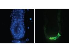
GFP Antibody - Immunofluorescence Microscopy of Anti-GFP (GOAT) Antibody. Tissue: E5.5 Hex-GFP transgenic mouse embryo. Primary antibody: Goat anti-GFP was used at 1:500 dilution. Secondary antibody: Fluorchrome conjugated Anti-goat IgG secondary antibody at 1:10,000 for 45 min at RT. Staining: GFP as green fluorescent signal with DAPI blue counterstain.
Select
GFP Goat anti-Aequorea victoria Polyclonal Antibody
Aequorea victoria
ELISA, IF, IHC, IP, WB
Unconjugated
1 mg/$715
Select
GFP Mouse Monoclonal (N/K) Antibody
Aequorea victoria
ELISA, IHC-Fr, IHC-P, WB
Unconjugated
0.1 ml/$662
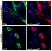
GFP Antibody - Confocal microscopy images of COS-7 cells transfected with expression constructs encoding membrane-tethered EGFP (membrane-EGFP; top) or nuclear Polycomb 2-EYFP fusion protein (Pc2-EYFP; bottom).  The natural fluorescence of the produced proteins is shown in the green channel (left), polyclonal anti-GFP antibody signal was detected in the red channel (right). The system was carefully tested for overlap of these two optical channels and images were scanned separately in sequential scanning mode. The blue nuclear stain is also shown.
Select
GFP Rabbit anti-Aequorea victoria Polyclonal (Polyclonal) Antibody
All species
ICC, IP, WB
Unconjugated
0.1 mg/$316
Select
GFP Goat anti-Aequorea victoria Polyclonal (Biotin) Antibody
Aequorea victoria
ELISA, IHC, WB
Biotin Conjugated
500 µg/$743
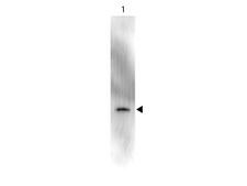
GFP Antibody - Anti-GFP Antibody - Western Blot. Western blot of GFP recombinant protein detected with polyclonal anti-GFP antibody. Lane 1 shows detection of a 33 kD band corresponding to a GFP containing recombinant protein (arrowhead) expressed in HeLa cells. Lane 2 shows no staining of a mock transfected HeLa cell lysate. A 4-12% Bis-Tris gradient gel was used for SDS-PAGE. The protein was transferred to nitrocellulose using standard methods. After blocking the membrane was probed with the primary antibody diluted to 1 ug/ml for 1 h at room temperature followed by washes and reaction with a 1:2500 dilution of IRDye 800 conjugated Donkey-a-Goat IgG [H&L] MX7 (. The IRDye 800 fluorescence image was captured using the Odyssey Infrared Imaging System developed by LI-COR. IRDye is a trademark of LI-COR, Inc. Other detection systems will yield similar results. This image was taken for the unconjugated form of this product. Other forms have not been tested.
Select
GFP Goat anti-Aequorea victoria Polyclonal (FITC) Antibody
Aequorea victoria
FLISA, IF, WB
FITC Conjugated
1 mg/$768
Select
GFP Mouse anti-Aequorea victoria Monoclonal Antibody
Aequorea victoria
ELISA, IHC, IHC-Fr, IHC-P, WB
Unconjugated
100 µl/$913
GFP Protein
Select
E. coli
None
Less than 1.0 EU/µg protein (determined by LAL method).
100 µg/$345; 500 µg/$657; 20 µg/$254
GFP ELISA Kit
Select
Sandwich
96-Well Strip Plate
All species
15.6 - 1000 pg/ml
Colorimetric - 450nm (TMB)
Cell Lysates
1 Plate/$632
GFP ELISA Kit
Select
Sandwich
96-Well Strip Plate
All species
1.56 - 100 ng/ml
Colorimetric - 450nm (TMB)
Biological Fluids
1 Plate/$831
GFP Antibody
Select
GFP Mouse anti-Mouse Monoclonal (1E4) Antibody
ICC, IHC, IP, WB
Unconjugated
100 µg/$400
GFP Antibody - Immuno-Fluorescence of Biotin Mouse anti-GFP antibody. Biotin mouse anti GFP used 1:5000 As referenced in:
Select
GFP Rabbit anti-Aequorea victoria Polyclonal (Biotin) Antibody
Aequorea victoria
ELISA, IF, IHC, WB
Biotin Conjugated
100 µg/$544
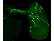
GFP Antibody - Immunofluorescence Microscopy of chicken anti-GFP antibody. Tissue: KruppleGAL4 driver line in Drosophila eye disc. Fixation: 0.5% PFA. Antigen retrieval: not required. Primary antibody: anti-GFP antibody diluted 1:500 for 2 hr at RT. Secondary antibody: Alexa™488 conjugated anti-Chicken IgG at 1:300 for 1 hr at RT.5% NGS in PBS with 0.1% Triton X-100 for 15 min. Localization: E2F-1 pS364 is nuclear and occasionally cytoplasmic. Staining: recombinant tau-myc-GFP protein as green fluorescent signal.
Select
GFP Chicken anti-Aequorea victoria Polyclonal Antibody
Aequorea victoria
ELISA, IF, IHC, WB
Unconjugated
100 µg/$462
Select
GFP Rabbit anti-Aequorea victoria Polyclonal Antibody
Aequorea victoria
ICC, IF, IHC, IHC-P, IP, WB
Unconjugated
50 µl/$771
GFP ELISA Kit
Select
Development Kit
Antibody Pair and Standard
All species
3.12 - 200 ng/ml
Colorimetric - 450nm (TMB)
Biological Fluids
500 Wells/$1,432; 1000 Wells/$2,163
GFP Antibody - Immunocytochemistry/Immunofluorescence analysis of GFP fusion gene transfeced HEK293 cells using THE TM GFP Antibody, pAb, Rabbit and Rabbit IgG Control (Whole Molecule), Purified The signal was developed with R-PE conjugated Goat Anti-Rabbit IgG.
Select
GFP Rabbit Monoclonal Antibody
Flo, IF, IP, WB
Unconjugated
100 µg/$572
GFP Antibody - Peroxidase - Western Blot. Western Blot showing detection of Green Fluorescent Protein (GFP). 50 ng of GFP (Lane 1) was run on a 4-20% gel and transferred to 0.45 micron nitrocellulose. After blocking with 1% BSA-TTBS (MB-013, diluted to 1X) 30 min at 20?, Anti-GFP (MOUSE) Monoclonal Antibody Peroxidase Conjugate Anti-GFP (MOUSE) Monoclonal Antibody Peroxidase Conjugate - 600-303-215 secondary antibody was used at 1:1000 in Blocking Buffer for Fluorescent Western Blot (p/n MB-070) and imaged using the Bio-Rad VersaDoc 4000 MP. Arrow indicates correct 28 kD molecular weight position expected for GFP. This image was taken for the unconjugated form of this product. Other forms have not been tested.
Select
GFP Mouse anti-Aequorea victoria Monoclonal (HRP) (9F9.F9) Antibody
Aequorea victoria
ELISA, IHC, WB
HRP Conjugated
1 mg/$947
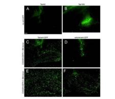
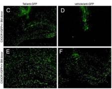
GFP Antibody - IHC - Immuno-Fluorescence of Biotin Mouse anti-GFP antibody. Biotin mouse anti GFP used 1:5000 As referenced in: Lu S-M, Tremblay M-E` , King IL, Qi J, Reynolds HM, et al. (2011) HIV-1 Tat-Induced Microgliosis and Synaptic Damage via Interactions between Peripheral and Central Myeloid Cells. PLoS ONE 6(9): e23915. doi:10.1371/journal.pone.0023915
Select
GFP Mouse anti-Aequorea victoria Monoclonal (Biotin) (9F9.F9) Antibody
Aequorea victoria
ELISA, IHC, WB
Biotin Conjugated
1 mg/$947
Select
GFP Mouse anti-Aequorea victoria Monoclonal (FITC) Antibody
Aequorea victoria
Flo, IF
FITC Conjugated
1 mg/$1,355
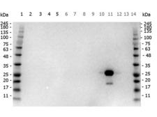
GFP Antibody - Western Blot of Goat anti-GFP antibody Peroxidase conjugated. Marker: Opal Pre-stained ladder  Lane 2: HEK293 lysate  Lane 3: HeLa Lysate  Lane 4: CHO/K1 Lysate  Lane 5: C2C12 Lysate. Lane 6: NIH/3T3 Lysate  Lane 7: Mouse Embryonic Fibroblast Lysate  Lane 8: E-coli HCP Control  Lane 9: 2 Epitope MBP Tag Marker Lysate  Lane 10: Recombinant Red Fluorescent Protein  Lane 11: Green Fluorescent Protein  Lane 12: Glutathione-S-Transferase Protein. Lane 13: Maltose Binding Protein  Marker: Opal Pre-stained ladder Load: 10 µg of lysate or 50ng of purified protein per lane. Primary antibody: goat anti-GFP peroxidase conjugated antibody at 1:1,000 overnight at 4C. Secondary antibody: none
Select
GFP Goat anti-Aequorea victoria Polyclonal (HRP) Antibody
Aequorea victoria
ELISA, IHC, WB
HRP Conjugated
1 mg/$749
Select
GFP Rabbit anti-Aequorea victoria Polyclonal (aa204-217) Antibody
Aequorea victoria
WB
Unconjugated
100 µg/$634
GFP Antibody
Select
GFP Rabbit Polyclonal (Biotin) Antibody
All species, Human
WB
Biotin Conjugated
100 µl/$321; 200 µl/$368; 500 µl/$526; 1 ml/$608; 2 ml/$839; 5 ml/$1,379
Viewing 1-25 of 120 product results


If you do not find the reagent or information you require, please contact Customer.Support@LSBio.com to inquire about additional products in development.

PLEASE NOTE

For RESEARCH USE ONLY. Intended for use by laboratory professionals. Not intended for human diagnostic or therapeutic purposes.

The data on this page has been compiled from LifeSpan internal sources, the National Center for Biotechnology Information (NCBI), and The Universal Protein Resource (UniProt).