Login
Registration enables users to use special features of this website, such as past
order histories, retained contact details for faster checkout, review submissions, and special promotions.
order histories, retained contact details for faster checkout, review submissions, and special promotions.
Forgot password?
Registration enables users to use special features of this website, such as past
order histories, retained contact details for faster checkout, review submissions, and special promotions.
order histories, retained contact details for faster checkout, review submissions, and special promotions.
Quick Order
Products
Antibodies
ELISA and Assay Kits
Research Areas
Infectious Disease
Resources
Purchasing
Reference Material
Contact Us
Location
Corporate Headquarters
Vector Laboratories, Inc.
6737 Mowry Ave
Newark, CA 94560
United States
Telephone Numbers
Customer Service: (800) 227-6666 / (650) 697-3600
Contact Us
Additional Contact Details
Login
Registration enables users to use special features of this website, such as past
order histories, retained contact details for faster checkout, review submissions, and special promotions.
order histories, retained contact details for faster checkout, review submissions, and special promotions.
Forgot password?
Registration enables users to use special features of this website, such as past
order histories, retained contact details for faster checkout, review submissions, and special promotions.
order histories, retained contact details for faster checkout, review submissions, and special promotions.
Quick Order
| Catalog Number | Size | Price |
|---|---|---|
| LS-C154219-100 | 100 µg (1.29 mg/ml) | $465 |
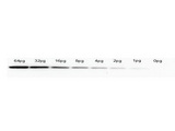
![GFP Antibody - Western blot of Rabbit anti-GFP antibody. Lane 1: Wild type GFP (0.1 g) was used to spike HeLa whole cell lysate. Lane 2: none. Load: 30 ug per lane. Primary antibody: GFP antibody at 1:1000 for overnight at 4C. Secondary antibody: IRDye800 Goat-a-Rabbit IgG [H&L] MX10 (611-132-122) at 1:10000 for 45 min at RT. Block: 5% BLOTTO in PBS overnight at 4C. Predicted/Observed size: 27 kDa for epitope tag GFP. Other band(s): none.](https://lsbio-7d62.kxcdn.com/image2/gfp-antibody-ls-c154219/136567_162184.jpg)
![GFP Antibody - Anti-GFP Antibody - Western Blot. Western blot of GFP protein detected with polyclonal anti-GFP antibody. Wild type GFP (0.1 ug) was used to spike 30 ug of a HeLa whole cell lysate. This antibody detects a 27 kD band corresponding to the epitope tag GFP. A 4-20% Tris-Glycine gradient gel was used for SDS-PAGE. The protein was transferred to nitrocellulose using standard methods. After blocking with 5% BLOTTO in PBS, the membrane was probed overnight at 4C with the primary antibody diluted in 5% BLOTTO to 1:1000, followed by washes and reaction with a 1:10000 dilution of IRDye 800 conjugated Goat-a-Rabbit IgG [H&L] MX10 (. IRDye 800 fluorescence image was captured using the Odyssey Infrared Imaging System developed by LI-COR. IRDye is a trademark of LI-COR, Inc. Other detection systems will yield similar results.](https://lsbio-7d62.kxcdn.com/image2/gfp-antibody-ls-c154219/81899_126473.jpg)

1 of 3
2 of 3
3 of 3
Polyclonal Rabbit anti‑Aequorea victoria GFP Antibody (IHC, IF, WB) LS‑C154219
Polyclonal Rabbit anti‑Aequorea victoria GFP Antibody (IHC, IF, WB) LS‑C154219
Antibody:
GFP Rabbit anti-Aequorea victoria Polyclonal Antibody
Application:
IHC, IF, WB, ELISA
Reactivity:
Aequorea victoria
Format:
Unconjugated, Unmodified
Toll Free North America
 (800) 227-6666
For Research Use Only
Overview
Antibody:
GFP Rabbit anti-Aequorea victoria Polyclonal Antibody
Application:
IHC, IF, WB, ELISA
Reactivity:
Aequorea victoria
Format:
Unconjugated, Unmodified
Specifications
Description
GFP antibody LS-C154219 is an unconjugated rabbit polyclonal antibody to aequorea victoria GFP. Validated for ELISA, IF, IHC and WB. Cited in 7 publications.
Host
Rabbit
Reactivity
Aequorea victoria
(tested or 100% immunogen sequence identity)
Clonality
IgG
Polyclonal
Conjugations
Unconjugated
Purification
Affinity chromatography
Modifications
Unmodified
Immunogen
The immunogen is a Green Fluorescent Protein (GFP) fusion protein corresponding to the full length amino acid sequence (246aa) derived from the jellyfish Aequorea victoria.
Specificity
Assay by immunoelectrophoresis resulted in a single precipitin arc against anti-Rabbit Serum and purified and partially purified Green Fluorescent Protein (Aequorea victoria). No reaction was observed against Human, Mouse or Rat serum proteins.
Applications
- IHC (1:200 - 1:3000)
- Immunofluorescence (1:500 - 1:5000)
- Western blot (1:500 - 1:5000)
- ELISA (1:20000 - 1:120000)

|
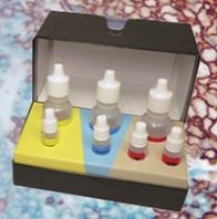
Performing IHC? See our complete line of Immunohistochemistry Reagents including antigen retrieval solutions, blocking agents
ABC Detection Kits and polymers, biotinylated secondary antibodies, substrates and more.
|
Usage
Anti-GFP antibody is designed to detect GFP and its variants such as rGFP, eGFP, S65T-GFP, RS-GFP, YFP, and EGFP. GFP antibody can be used to detect GFP by ELISA (sandwich or capture) for the direct binding of antigen and recognizes wild type, recombinant and enhanced forms of GFP. Biotin conjugated polyclonal anti-GFP used in a sandwich ELISA is well suited to titrate GFP in solution using this antibody in combination with a monoclonal anti-GFP antibody using either form of the antibody as the capture or detection antibodies. However, use the monoclonal form only for the detection of wild type or recombinant GFP as this form does not sufficiently detect 'enhanced' GFP. The detection antibody is typically conjugated to biotin and subsequently reacted with streptavidin conjugated HRP. Fluorochrome conjugated polyclonal anti-GFP can be used to detect GFP by immunofluorescence microscopy in prokaryotic (E. coli) and eukaryotic (CHO cells) expression systems and can detect GFP containing inserts. Significant amplification of signal is achieved using fluorochrome conjugated polyclonal anti-GFP relative to the fluorescence of GFP alone. For immunoblotting use either alkaline phosphatase or peroxidase conjugated polyclonal anti-GFP to detect GFP or GFP containing proteins on western blots. Optimal titers for applications should be determined by the researcher.
Presentation
0.02 M Potassium Phosphate, pH 7.2, 0.15 M NaCl, 0.01% Sodium Azide
Storage
Short term: store at 4°C. Long term: aliquot and store at -20°C. Avoid freeze-thaw cycles.
Restrictions
For research use only. Intended for use by laboratory professionals.
LSBio Ratings
GFP Antibody for IHC, IF/Immunofluorescence, WB/Western, ELISA LS-C154219 has an LSBio Rating of
Publications (4.6)
Learn more about The LSBio Ratings Algorithm
Publications (7)
Splice variants of the dual specificity tyrosine phosphorylation-regulated kinase 4 (DYRK4) differ in their subcellular localization and catalytic activity. Papadopoulos C, Arato K, Lilienthal E, Zerweck J, Schutkowski M, Chatain N, Mller-Newen G, Becker W, de la Luna S. The Journal of biological chemistry. 2011 286:5494-505.
The syntaxin 4 N terminus regulates its basolateral targeting by munc18c-dependent and -independent mechanisms. Torres J, Funk HM, Zegers MM, ter Beest MB. The Journal of biological chemistry. 2011 286:10834-46.
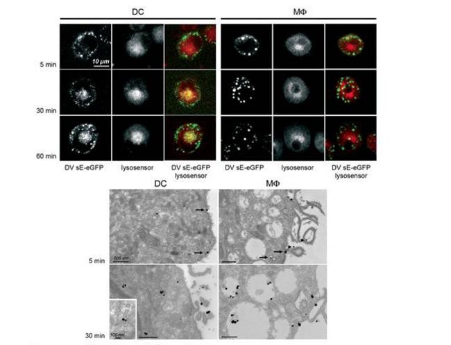
Vaccinia extracellular virions enter cells by macropinocytosis and acid-activated membrane rupture. Schmidt FI, Bleck CK, Helenius A, Mercer J. The EMBO journal. 2011 30:3647-61.
Retinoblastoma tumor-suppressor protein phosphorylation and inactivation depend on direct interaction with Pin1. Rizzolio F, Lucchetti C, Caligiuri I, Marchesi I, Caputo M, Klein-Szanto AJ, Bagella L, Castronovo M, Giordano A. Cell death and differentiation. 2012 19:1152-61.
High-throughput, multiplexed analysis of 3-nitrotyrosine in individual proteins. Jin H, Zangar RC. Current protocols in toxicology / editorial board, Mahin D. Maines (editor-in-chief) ... [et al.]. 2012 Unit 17.15.
Hepatocellular Carcinoma Cell-Secreted Exosomal MicroRNA-210 Promotes Angiogenesis In Vitro and In Vivo. Lin XJ, Fang JH, Yang XJ, Zhang C, Yuan Y, Zheng L, Zhuang SM. Molecular therapy. Nucleic acids. 2018 June;11:243-252. (Aequorea victoria)
Fibrotic microenvironment promotes the metastatic seeding of tumor cells via activating the fibronectin 1/secreted phosphoprotein 1-integrin signaling. Zhang C, Wu M, Zhang L, Shang LR, Fang JH, Zhuang SM. Oncotarget. 2016 July;7:45702-45714. (Aequorea victoria)
Customer Reviews (0)
Featured Products
Species:
Aequorea victoria
Applications:
IHC, IHC - Paraffin, Western blot, Immunoprecipitation, ELISA
Species:
Aequorea victoria
Applications:
IHC, IHC - Paraffin, IHC - Frozen, Western blot, ELISA
Species:
Aequorea victoria
Applications:
IHC, Immunofluorescence, Western blot, ELISA
Species:
Aequorea victoria
Applications:
IHC, Immunofluorescence, Western blot, Flow Cytometry, ELISA
Species:
Aequorea victoria, All species
Applications:
ICC, Western blot, Immunoprecipitation
Request SDS/MSDS
To request an SDS/MSDS form for this product, please contact our Technical Support department at:
Technical.Support@LSBio.com
Requested From: United States
Date Requested: 3/12/2025
Date Requested: 3/12/2025


