Products
Research Areas
COVID-19
Resources
Login
Quick Order
Cart Cart lightblue
Login
Registration enables users to use special features of this website, such as past
order histories, retained contact details for faster checkout, review submissions, and special promotions.


Fields marked with a * are required.

Login
Quick Order
Contact Us

Location


Corporate Headquarters

Vector Laboratories, Inc.
6737 Mowry Ave
Newark, CA 94560
United States

Telephone Numbers


Customer Service: (800) 227-6666 / (650) 697-3600


Contact Us


Additional Contact Details

Login
Registration enables users to use special features of this website, such as past
order histories, retained contact details for faster checkout, review submissions, and special promotions.


Fields marked with a * are required.

Login
Quick Order

MDM2

MDM2 oncogene, E3 ubiquitin protein ligase

E3 ubiquitin-protein ligase that mediates ubiquitination of p53/TP53, leading to its degradation by the proteasome. Inhibits p53/TP53- and p73/TP73-mediated cell cycle arrest and apoptosis by binding its transcriptional activation domain. Also acts as a ubiquitin ligase E3 toward itself and ARRB1. Permits the nuclear export of p53/TP53. Promotes proteasome-dependent ubiquitin-independent degradation of retinoblastoma RB1 protein. Inhibits DAXX-mediated apoptosis by inducing its ubiquitination and degradation. Component of the TRIM28/KAP1-MDM2-p53/TP53 complex involved in stabilizing p53/TP53. Also component of the TRIM28/KAP1-ERBB4-MDM2 complex which links growth factor and DNA damage response pathways. Mediates ubiquitination and subsequent proteasome degradation of DYRK2 in nucleus. Ubiquitinates IGF1R and SNAI1 and promotes them to proteasomal degradation.

Gene Name: MDM2 oncogene, E3 ubiquitin protein ligase
Synonyms: MDM2, ACTFS, Double minute 2 protein, Hdm2, MDM2 variant FB30, HDMX, Oncoprotein Mdm2, p53-binding protein Mdm2, MDM2 variant FB28
Target Sequences: NM_002392 NP_002383.2 Q00987

Publications (3)

1
Stabilization of HIF-1a and HIF-2a, up-regulation of MYCC and accumulation of stabilized p53 constitute hallmarks of CNS-PNET animal model. Malchenko S, Sredni ST, Bi Y, Margaryan NV, Boyineni J, Mohanam I, Tomita T, Davuluri RV, Soares MB. PloS one. 2017 12:e0173106. (IHC, WB; Mouse) [Full Text Article] [PubMed:28249000] [PMC:PMC5332108] Related Antibodies: LS-C199239.
2
Characterization of brain tumor initiating cells isolated from an animal model of CNS primitive neuroectodermal tumors. Malchenko S, Sredni ST, Boyineni J, Bi Y, Margaryan NV, Guda MR, Kostenko Y, Tomita T, Davuluri RV, Velpula K, Hendrix MJC, Soares MB. Oncotarget. 2018 February;9:13733-13747. (Human) [Full Text Article] [PubMed:29568390] [PMC:PMC5862611] Related Antibodies: LS-C199239.
3
Targeting the TP53/MDM2 axis enhances radiation sensitivity in atypical teratoid rhabdoid tumors. Irina Alimova, Dong Wang, Etienne Danis, Angela Pierce, Andrew Donson, Natalie Serkova, Krishna Madhavan, Senthilnath Lakshmanachetty, Ilango Balakrishnan, Nicholas K Foreman, Siddhartha Mitra, Sujatha Venkataraman, Rajeev Vibhakar. International journal of oncology. 2022 March;60:32. [Full Text Article] [PubMed:35179215] [PMC:PMC8878716] Related Antibodies: LS-C199239.

☰ Filters
Products
Antibodies (158)
Primary (158)
MDM2 (158)
Human (153)
Mouse (54)
Rat (31)
Bovine (1)
Dog (2)
Gibbon (1)
Horse (1)
Monkey (9)
Orangutan (1)
Rabbit (1)
IHC (74)
IHC-Fr (4)
IHC-P (28)
WB (120)
DB (14)
ELISA (50)
ICC (5)
IF (27)
IHC-R (1)
IP (10)
Peptide-ELISA (8)
rabbit (101)
mouse (56)
goat (1)
Product Group
IHCPlus (3)
IgG (61)
IgG1 (40)
IgG1,k (6)
IgG2a (4)
IgG2b (4)
IgG2b,k (1)
monoclonal mc (56)
polyclonal pc (102)
1806CT309.52.1 (1)
1A7 (1)
MDM2/2414 (3)
OTI15C5 (2)
OTI15F3 (2)
OTI16H3 (2)
OTI17B3 (2)
OTI1B4 (2)
OTI1E5 (2)
OTI1E6 (2)
OTI20D1 (2)
OTI22D6 (2)
OTI22G3 (2)
OTI24C7 (2)
OTI25B9 (2)
OTI2H4 (2)
OTI3F1 (1)
OTI4B3 (2)
OTI4E5 (2)
OTI5E6 (2)
OTI6H3 (2)
OTI7F7 (2)
OTI8C6 (2)
OTI8F7 (2)
SMP14 (2)
AP Conjugated (1)
APC (1)
APC Conjugated (7)
Atto 390 Conjugated (1)
Atto 488 Conjugated (1)
Atto 594 Conjugated (1)
Azide-free (5)
Biotin Conjugated (9)
BSA-free (1)
Carrier-free (20)
Cy3 Conjugated (1)
Cy7 Conjugated (1)
FITC Conjugated (9)
HRP Conjugated (8)
Magnetic beads Conjugated (1)
PE Conjugated (7)
PerCP Conjugated (1)
Ready-to-Use (2)
RPE Conjugated (1)
Sepharose Conjugated (1)
Unconjugated (108)
aa119-438 (38)
pSer166 (9)
aa1-321 (8)
Ser166 (7)
aa393-424 (7)
pSer395 (7)
pThr218 (7)
Internal (5)
110-190 aa (2)
120-200 aa (2)
C-Terminus (2)
aa103-119 (2)
360-440 aa, C-terminal (1)
381-430 aa, C-terminal (1)
Ab-186/188 (1)
Phe414 (1)
Ser160 (1)
Tyr394 (1)
aa132-181 (1)
aa151-200 (1)
aa154-167 (1)
aa177-195 (1)
aa179-193 (1)
aa26-169 (1)
aa343-358 (1)
aa391-440 (1)
aa418-432 (1)
p166 (1)
pSer185 (1)
pSer186 (1)
pSer186+Ser188 (1)
pSer260 (1)
pTyr394 (1)
No (157)
Yes (1)
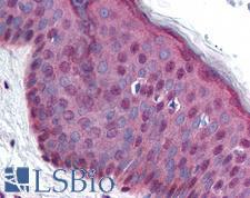
MDM2 Antibody - Anti-MDM2 antibody IHC of human skin. Immunohistochemistry of formalin-fixed, paraffin-embedded tissue after heat-induced antigen retrieval. Antibody concentration 10 ug/ml.
Select
MDM2 Mouse anti-Human Monoclonal (aa154-167) (SMP14) Antibody
Human
IHC, IHC-Fr, IHC-P, IHC-R
Unconjugated
50 µg/$460
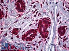
MDM2 Antibody - Anti-MDM2 antibody IHC of human breast. Immunohistochemistry of formalin-fixed, paraffin-embedded tissue after heat-induced antigen retrieval. Antibody concentration 2.5 ug/ml.
Select
MDM2 Mouse anti-Human Monoclonal (1A7) Antibody
Human
ELISA, IHC, IHC-P, WB
Unconjugated
50 µg/$460
Select
MDM2 Rabbit anti-Mouse Polyclonal (aa177-195) Antibody
Mouse
ELISA, WB
Unconjugated
50 µg/$448
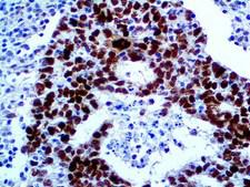
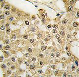
MDM2 Antibody - IHC of MDM2 on an FFPE Testicular Cancer Tissue
Select
MDM2 Mouse anti-Human Monoclonal Antibody
Human
IHC-Fr, IHC-P
Unconjugated
0.1 ml/$340
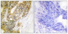
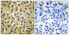
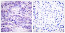
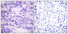
MDM2 Antibody - Immunohistochemistry analysis of paraffin-embedded human colon carcinoma tissue, using MDM2 Antibody. The picture on the right is blocked with the synthesized peptide.
Select
MDM2 Rabbit anti-Human Polyclonal (aa151-200) Antibody
Human
IF, IHC, IHC-P, Peptide-ELISA
Unconjugated
50 µl/$334; 100 µl/$397
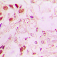
MDM2 Antibody - Immunohistochemical analysis of MDM2 staining in human lung cancer formalin fixed paraffin embedded tissue section. The section was pre-treated using heat mediated antigen retrieval with sodium citrate buffer (pH 6.0). The section was then incubated with the antibody at room temperature and detected using an HRP conjugated compact polymer system. DAB was used as the chromogen. The section was then counterstained with hematoxylin and mounted with DPX.
Select
MDM2 Rabbit anti-Human Polyclonal (C-Terminus) Antibody
Human, Monkey
ICC, IF, IHC, IHC-P, WB
Unconjugated
100 µl/$299
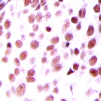
MDM2 Antibody - Immunohistochemical analysis of MDM2 staining in human prostate cancer formalin fixed paraffin embedded tissue section. The section was pre-treated using heat mediated antigen retrieval with sodium citrate buffer (pH 6.0). The section was then incubated with the antibody at room temperature and detected using an HRP polymer system. DAB was used as the chromogen. The section was then counterstained with hematoxylin and mounted with DPX.
Select
MDM2 Rabbit anti-Human Polyclonal (Internal) Antibody
Rat, Human, Monkey
ICC, IF, IHC, IHC-P, IP, WB
Unconjugated
100 µl/$321
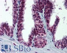
MDM2 Antibody - Anti-MDM2 antibody IHC of human prostate. Immunohistochemistry of formalin-fixed, paraffin-embedded tissue after heat-induced antigen retrieval. Antibody concentration 5 ug/ml.
Select
MDM2 Rabbit anti-Human Polyclonal (pSer395) Antibody
Human
DB, IHC, IHC-P
Unconjugated
200 µl/$440
Select
MDM2 Mouse anti-Human Monoclonal (aa26-169) Antibody
Human
IF, IHC, IHC-Fr, IHC-P, IP, WB
Unconjugated
50 µg/$778
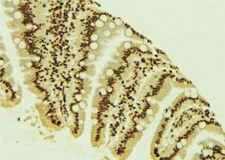
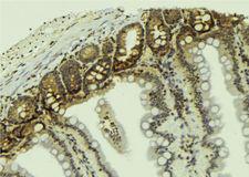
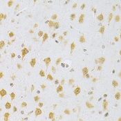
MDM2 Antibody - 1:100 staining mouse colon tissue by IHC-P. The sample was formaldehyde fixed and a heat mediated antigen retrieval step in citrate buffer was performed. The sample was then blocked and incubated with the antibody for 1.5 hours at 22°C. An HRP conjugated goat anti-rabbit antibody was used as the secondary.
Select
MDM2 Rabbit anti-Human Polyclonal Antibody
Mouse, Rat, Human, Monkey
IF, IHC, Peptide-ELISA, WB
Unconjugated
100 µl/$379; 200 µl/$421
MDM2 Antibody - Anti-MDM2 Antibody - Western Blot. Western blot using affinity purified Anti-MDM2 (Rabbit) is shown to detect a band (arrow) corresponding to mouse MDM2 protein present in mouse MEF cells (lane 2) but not human kidney HEK293 cells (lane 1). Approximately 35 ug of lysate was separated by 4-20% Tris Glycine SDS-PAGE. After blocking the membrane with 5% normal goat serum, 0.5% BLOTTO in PBS, the membrane was probed for overnight at 4° with the primary antibody diluted to 1:500 in 1% normal goat serum, 0.1% BLOTTO in PBS. The membrane was washed and reacted with a 1:10000 dilution of IRDye800 conjugated Gt-a-Rabbit IgG [H&L] ( for 45 min at room temperature (800 nm channel, green). Molecular weight estimation was made by comparison to prestained MW markers indicated at the right (700 nm channel, red). IRDye800 fluorescence image was captured using the Ddyssey Infrared Imaging System developed by LI-COR. IRDye is a trademark of LI-COR, Inc. Other detection systems will yield similar results.
Select
MDM2 Rabbit anti-Mouse Polyclonal Antibody
Mouse
ELISA, IHC, IHC-P, IP, WB
Unconjugated
100 µg/$567
Select
MDM2 Mouse anti-Human Monoclonal (PE) Antibody
Mouse, Human
IHC, IP, WB
PE Conjugated
100 µl/$848
Select
MDM2 Mouse anti-Human Monoclonal (Tyr394) Antibody
Mouse, Human
ELISA, WB
Unconjugated
50 µg/$295; 100 µg/$335; 200 µg/$394
MDM2 Antibody - Antibody (0.3 ug/ml) staining of Human Liver lysate (35 ug protein in RIPA buffer). Primary incubation was 1 hour. Detected by chemiluminescence.
Select
MDM2 Goat anti-Human Polyclonal (aa179-193) Antibody
Human, Monkey
Peptide-ELISA, WB
Unconjugated
100 µg/$503
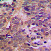
MDM2 Antibody - Immunohistochemical analysis of MDM2 (pS166) staining in human breast cancer formalin fixed paraffin embedded tissue section. The section was pre-treated using heat mediated antigen retrieval with sodium citrate buffer (pH 6.0). The section was then incubated with the antibody at room temperature and detected using an HRP conjugated compact polymer system. DAB was used as the chromogen. The section was then counterstained with hematoxylin and mounted with DPX.
Select
MDM2 Rabbit anti-Human Polyclonal (pSer166) Antibody
Human
ICC, IF, IHC, IHC-P, IP, WB
Unconjugated
100 µl/$315
MDM2 Antibody - 1:100 staining mouse colon tissue by IHC-P. The sample was formaldehyde fixed and a heat mediated antigen retrieval step in citrate buffer was performed. The sample was then blocked and incubated with the antibody for 1.5 hours at 22°C. An HRP conjugated goat anti-rabbit antibody was used as the secondary.
Select
MDM2 Rabbit anti-Human Polyclonal (pSer166) Antibody
Mouse, Human, Monkey
IF, IHC, Peptide-ELISA, WB
Unconjugated
100 µl/$379; 200 µl/$421
Select
MDM2 Rabbit anti-Human Polyclonal (aa103-119) Antibody
Rat, Human
WB
Unconjugated
100 µg/$380
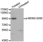
MDM2 Antibody - Western blot analysis of extracts of 293 cells.
Select
MDM2 Rabbit anti-Human Polyclonal (pSer260) Antibody
WB
Unconjugated
20 µl/$287; 100 µl/$431
MDM2 Antibody - Peptide - + Immunohistochemical analysis of paraffin-embedded human breast carcinoma tissue using MDM2 antibody.
Select
MDM2 Rabbit anti-Human Polyclonal Antibody
Rat, Human
ELISA, IF, IHC, WB
Unconjugated
100 µg/$357
MDM2 Antibody - P-peptide - + Immunohistochemistry analysis of paraffin-embedded human breast carcinoma tissue using MDM2 (Phospho-Ser166) antibody.
Select
MDM2 Rabbit anti-Human Polyclonal (pSer166) Antibody
Mouse, Human
ELISA, IF, IHC, WB
Unconjugated
100 µg/$357
MDM2 Antibody - Western blot of MDM2 phosphorylation expression in COS7 whole cell lysates,The lane on the left is treated with the antigen-specific peptide.
Select
MDM2 Rabbit anti-Human Polyclonal (pSer166) Antibody
Mouse, Human
ICC, IF, IHC, WB
Unconjugated
50 µg/$305; 100 µg/$357
MDM2 Antibody - Formalin-fixed and paraffin-embedded human breast carcinoma tissue reacted with the MDM2 Antibody (S166) , which was peroxidase-conjugated to the secondary antibody, followed by DAB staining. This data demonstrates the use of this antibody for immunohistochemistry; clinical relevance has not been evaluated.
Select
MDM2 Rabbit anti-Human Polyclonal (Ser166) Antibody
Human
ICC, IF, IHC, IHC-P, WB
Unconjugated
400 µl/$393
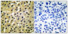
MDM2 Antibody - Immunohistochemistry analysis of paraffin-embedded human breast carcinoma tissue, using MDM2 Antibody. The picture on the right is blocked with the synthesized peptide.
Select
MDM2 Rabbit anti-Human Polyclonal (aa391-440) Antibody
Mouse, Human
IF, IHC, IHC-P, Peptide-ELISA, WB
Unconjugated
50 µl/$334; 100 µl/$397
Select
MDM2 Rabbit anti-Human Polyclonal (110-190 aa) Antibody
Mouse, Human, Monkey
ELISA, IF, IHC, WB
Unconjugated
50 µg/$295; 100 µg/$335; 200 µg/$394
MDM2 Antibody - Immunohistochemistry analysis of paraffin-embedded human breast carcinoma, using MDM2 (Phospho-Ser166) Antibody. The picture on the right is blocked with the phospho peptide.
Select
MDM2 Rabbit anti-Human Polyclonal (pSer166) Antibody
Mouse, Human
IF, IHC, IHC-P, Peptide-ELISA, WB
Unconjugated
50 µl/$334; 100 µl/$397
Viewing 1-25 of 158 product results


Filtered By:
Products: Antibodies


If you do not find the reagent or information you require, please contact Customer.Support@LSBio.com to inquire about additional products in development.

PLEASE NOTE

For RESEARCH USE ONLY. Intended for use by laboratory professionals. Not intended for human diagnostic or therapeutic purposes.

The data on this page has been compiled from LifeSpan internal sources, the National Center for Biotechnology Information (NCBI), and The Universal Protein Resource (UniProt).