Login
Registration enables users to use special features of this website, such as past
order histories, retained contact details for faster checkout, review submissions, and special promotions.
order histories, retained contact details for faster checkout, review submissions, and special promotions.
Forgot password?
Registration enables users to use special features of this website, such as past
order histories, retained contact details for faster checkout, review submissions, and special promotions.
order histories, retained contact details for faster checkout, review submissions, and special promotions.
Quick Order
Products
Antibodies
ELISA and Assay Kits
Research Areas
Infectious Disease
Resources
Purchasing
Reference Material
Contact Us
Location
Corporate Headquarters
Vector Laboratories, Inc.
6737 Mowry Ave
Newark, CA 94560
United States
Telephone Numbers
Customer Service: (800) 227-6666 / (650) 697-3600
Contact Us
Additional Contact Details
Login
Registration enables users to use special features of this website, such as past
order histories, retained contact details for faster checkout, review submissions, and special promotions.
order histories, retained contact details for faster checkout, review submissions, and special promotions.
Forgot password?
Registration enables users to use special features of this website, such as past
order histories, retained contact details for faster checkout, review submissions, and special promotions.
order histories, retained contact details for faster checkout, review submissions, and special promotions.
Quick Order
| Catalog Number | Size | Price |
|---|---|---|
| LS-C369208-50 | 50 µg | $294 |
| LS-C369208-100 | 100 µg | $360 |
1 of 4
2 of 4
3 of 4
4 of 4
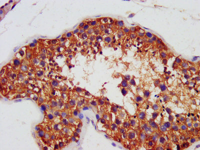
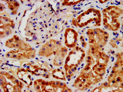
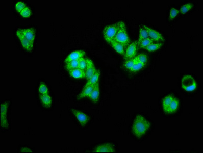
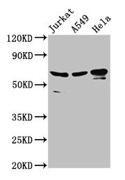
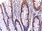
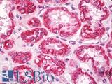
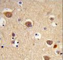
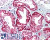
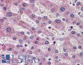
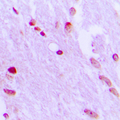
Polyclonal Rabbit anti‑Human AIFM1 / AIF / PDCD8 Antibody (aa103‑612) LS‑C369208
Polyclonal Rabbit anti‑Human AIFM1 / AIF / PDCD8 Antibody (aa103‑612) LS‑C369208
Toll Free North America
 (800) 227-6666
For Research Use Only
Overview
Specifications
Description
PDCD8 antibody LS-C369208 is an unconjugated rabbit polyclonal antibody to human PDCD8 (AIFM1 / AIF) (aa103-612). Validated for ELISA.
Target
Human AIFM1 / AIF / PDCD8
Synonyms
AIFM1 | AIF | COXPD6 | PDCD8 | Programmed cell death 8
Host
Rabbit
Reactivity
Human
(tested or 100% immunogen sequence identity)
Clonality
IgG
Polyclonal
Purification
Caprylic acid and ammonium sulfate precipitation
Modifications
Unmodified
Immunogen
Recombinant human Apoptosis-inducing factor 1, mitochondrial protein (aa103-612).
Epitope
aa103-612
Specificity
Human AIFM1 / AIF / PDCD8
Applications
- ELISA
Presentation
PBS, pH 7.4, 0.03% Proclin 300, 50% glycerol.
Storage
Short term: -20°C; Long term: -80°C; Avoid freeze-thaw cycles.
Restrictions
For research use only. Intended for use by laboratory professionals.
About AIFM1 / AIF / PDCD8
Publications (0)
Customer Reviews (0)
Featured Products
Species:
Human, Monkey, Mouse, Rat, Bat, Bovine, Dog, Hamster, Horse, Pig, Rabbit
Applications:
IHC, IHC - Paraffin, IHC - Frozen, Western blot, Immunoprecipitation
Species:
Human, Mouse, Rat
Applications:
IHC, IHC - Paraffin, Western blot
Species:
Human
Applications:
IHC, IHC - Paraffin, Immunofluorescence, Western blot, Flow Cytometry
Species:
Human, Mouse
Applications:
IHC, IHC - Paraffin, Western blot
Species:
Human, Mouse, Rat
Applications:
IHC, IHC - Paraffin, ICC, Western blot, ELISA
Species:
Human, Mouse, Rat, Sheep
Applications:
IHC, IHC - Paraffin, ICC, Immunofluorescence, Western blot, Immunoprecipitation
Request SDS/MSDS
To request an SDS/MSDS form for this product, please contact our Technical Support department at:
Technical.Support@LSBio.com
Requested From: United States
Date Requested: 1/19/2025
Date Requested: 1/19/2025


