Products
Research Areas
COVID-19
Resources
Login
Quick Order
Cart Cart lightblue
Login
Registration enables users to use special features of this website, such as past
order histories, retained contact details for faster checkout, review submissions, and special promotions.


Fields marked with a * are required.

Login
Quick Order
Contact Us

Location


Corporate Headquarters

Vector Laboratories, Inc.
6737 Mowry Ave
Newark, CA 94560
United States

Telephone Numbers


Customer Service: (800) 227-6666 / (650) 697-3600


Contact Us


Additional Contact Details

Login
Registration enables users to use special features of this website, such as past
order histories, retained contact details for faster checkout, review submissions, and special promotions.


Fields marked with a * are required.

Login
Quick Order

ULK1

unc-51 like autophagy activating kinase 1

Serine/threonine-protein kinase involved in autophagy in response to starvation. Acts upstream of phosphatidylinositol 3-kinase PIK3C3 to regulate the formation of autophagophores, the precursors of autophagosomes. Part of regulatory feedback loops in autophagy: acts both as a downstream effector and negative regulator of mammalian target of rapamycin complex 1 (mTORC1) via interaction with RPTOR. Activated via phosphorylation by AMPK and also acts as a regulator of AMPK by mediating phosphorylation of AMPK subunits PRKAA1, PRKAB2 and PRKAG1, leading to negatively regulate AMPK activity. May phosphorylate ATG13/KIAA0652 and RPTOR; however such data need additional evidences. Plays a role early in neuronal differentiation and is required for granule cell axon formation.

Gene Name: unc-51 like autophagy activating kinase 1
Family/Subfamily: Protein Kinase , ULK
Synonyms: ULK1, ATG1A, ATG1, HATG1, KIAA0722, Unc-51-like kinase 1, Unc51.1, UNC51
Target Sequences: NM_003565 NP_003556.1 O75385

Publications (1)

1
The signaling pathways implicated in impairment of hepatic autophagy in glycogen storage disease type Ia. Sudeep Gautam, Lisa Zhang, Irina Arnaoutova, Cheol Lee, Brian C Mansfield, Janice Y Chou. Human molecular genetics. 2020 March;29:834-844. [Full Text Article] [PubMed:31961433] [PMC:PMC7104680]

☰ Filters
Products
Antibodies (89)
Primary (89)
ULK1 (89)
Human (89)
Mouse (23)
Rat (20)
Bovine (3)
Chicken (3)
Chimpanzee (3)
Dog (3)
Gibbon (3)
Guinea pig (3)
Hamster (3)
Horse (3)
Insect (3)
Monkey (4)
Orangutan (3)
Pig (3)
Rabbit (3)
Xenopus (3)
Zebrafish (3)
IHC (10)
IHC-P (3)
WB (55)
Flo (1)
DB (28)
ELISA (56)
IF (14)
IP (1)
Peptide-ELISA (2)
rabbit (87)
mouse (1)
sheep (1)
Product Group
IHCPlus (2)
IgG (67)
IgG1,k (1)
monoclonal mc (2)
polyclonal pc (87)
2H8 (1)
D1H4 (1)
APC Conjugated (9)
Atto 390 Conjugated (1)
Atto 488 Conjugated (1)
Atto 594 Conjugated (1)
Azide-free (8)
Biotin Conjugated (11)
FITC Conjugated (11)
HRP Conjugated (10)
PE Conjugated (8)
PerCP Conjugated (1)
RPE Conjugated (1)
Unconjugated (35)
pSer556 (15)
aa296-322 (7)
aa538-564 (7)
pSer317 (7)
pSer467 (7)
aa642-672 (6)
Internal (5)
aa190-239 (4)
aa350-410 (2)
pSer555 (2)
C-terminal (1)
Ser638 (1)
Ser757 (1)
aa400-499 (1)
aa542-565 (1)
aa670-700 (1)
pSer757 (1)
No (89)
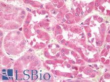
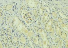
ULK1 Antibody - Anti-ULK1 antibody IHC staining of human kidney. Immunohistochemistry of formalin-fixed, paraffin-embedded tissue after heat-induced antigen retrieval. Antibody concentration 10 ug/ml.
Select
ULK1 Rabbit anti-Human Polyclonal Antibody
Mouse, Rat, Human
ELISA, IF, IHC, IHC-P, WB
Unconjugated
0.05 mg/$460
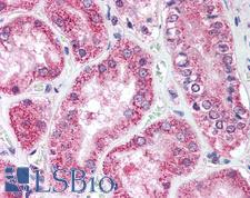
ULK1 Antibody - Anti-ULK1 antibody IHC of human kidney. Immunohistochemistry of formalin-fixed, paraffin-embedded tissue after heat-induced antigen retrieval. Antibody concentration 10 ug/ml.
Select
ULK1 Rabbit anti-Human Polyclonal Antibody
Human, Monkey
IHC, IHC-P, WB
Unconjugated
50 µg/$460
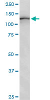
ULK1 Antibody - ULK1 monoclonal antibody (M01), clone 2H8. Western Blot analysis of ULK1 expression in human colon.
Select
ULK1 Mouse anti-Human Monoclonal (2H8) Antibody
Human
ELISA, WB
Unconjugated
100 µg/$423
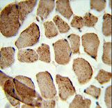
ULK1 Antibody - Formalin-fixed and paraffin-embedded human skeletal muscle reacted with Denatured ATG1 Antibody, which was peroxidase-conjugated to the secondary antibody, followed by DAB staining. This data demonstrates the use of this antibody for immunohistochemistry; clinical relevance has not been evaluated.
Select
ULK1 Rabbit anti-Human Polyclonal Antibody
Mouse, Human
Flo, IHC, IHC-P, WB
Unconjugated
400 µl/$393
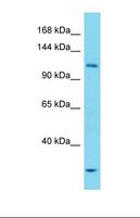
ULK1 Antibody - Western blot of Human Thymus Tumor. ULK1 antibody dilution 1.0 ug/ml.  This image was taken for the unconjugated form of this product. Other forms have not been tested.
Select
ULK1 Rabbit anti-Human Polyclonal (aa190-239) Antibody
Human
WB
Unconjugated
100 µl/$424
Select
ULK1 Sheep anti-Human Polyclonal (aa350-410) Antibody
Mouse, Rat, Human
IHC, WB
Unconjugated
500 µg/$393
Select
ULK1 Rabbit anti-Human Monoclonal (pSer555) (D1H4) Antibody
Mouse, Rat, Human
IP, WB
Unconjugated
100 µl/$689
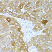
ULK1 Antibody - Immunohistochemistry of paraffin-embedded Mouse liver using ULk1 Polyclonal Antibody at dilution of 1:100 (40x lens).
Select
ULK1 Rabbit anti-Human Polyclonal Antibody
Mouse, Rat, Human
IF, IHC
Unconjugated
60 µl/$331; 120 µl/$403; 200 µl/$529
Select
ULK1 Rabbit anti-Human Polyclonal Antibody
Human
ELISA, IHC, WB
Unconjugated
100 µg/$351
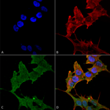
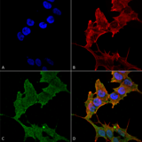
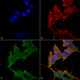
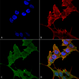
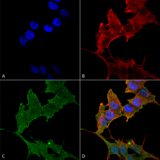
ULK1 Antibody - Immunocytochemistry/Immunofluorescence analysis using Rabbit Anti-ULK1 Polyclonal Antibody. Tissue: Neuroblastoma cell line (SK-N-BE). Species: Human. Fixation: 4% Formaldehyde for 15 min at RT. Primary Antibody: Rabbit Anti-ULK1 Polyclonal Antibody  at 1:100 for 60 min at RT. Secondary Antibody: Goat Anti-Rabbit ATTO 488 at 1:200 for 60 min at RT. Counterstain: Phalloidin Texas Red F-Actin stain; DAPI (blue) nuclear stain at 1:1000, 1:5000 for 60 min at RT, 5 min at RT. Localization: Cytoplasm, Preautophagosomal Structure. Magnification: 60X. (A) DAPI (blue) nuclear stain (B) Phalloidin Texas Red F-Actin stain (C) ULK1 Antibody (D) Composite.
Select
ULK1 Rabbit anti-Human Polyclonal Antibody
Mouse, Rat, Human
IF, WB
Unconjugated
100 µg/$442
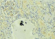
ULK1 Antibody - 1:100 staining mouse kidney tissue by IHC-P. The sample was formaldehyde fixed and a heat mediated antigen retrieval step in citrate buffer was performed. The sample was then blocked and incubated with the antibody for 1.5 hours at 22°C. An HRP conjugated goat anti-rabbit antibody was used as the secondary.
Select
ULK1 Rabbit anti-Human Polyclonal (pSer556) Antibody
Mouse, Rat, Human
IHC, Peptide-ELISA, WB
Unconjugated
100 µl/$379; 200 µl/$421
ULK1 Antibody - 1:100 staining mouse kidney tissue by IHC-P. The sample was formaldehyde fixed and a heat mediated antigen retrieval step in citrate buffer was performed. The sample was then blocked and incubated with the antibody for 1.5 hours at 22°C. An HRP conjugated goat anti-rabbit antibody was used as the secondary.
Select
ULK1 Rabbit anti-Human Polyclonal (aa542-565) Antibody
Mouse, Rat, Human
IHC, Peptide-ELISA, WB
Unconjugated
100 µl/$379; 200 µl/$421
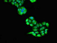
ULK1 Antibody - Immunofluorescent analysis of PC3 cells diluted at 1:100 and Alexa Fluor 488-congugated AffiniPure Goat Anti-Rabbit IgG(H+L)
Select
ULK1 Rabbit anti-Human Polyclonal Antibody
Human
ELISA, IF
Unconjugated
50 µg/$294; 100 µg/$360
Select
ULK1 Rabbit anti-Human Polyclonal (C-terminal) Antibody
Mouse, Human
ELISA, IHC
Unconjugated
50 µg/$295; 100 µg/$335; 200 µg/$394
Select
ULK1 Rabbit anti-Human Polyclonal (aa350-410) Antibody
Mouse, Rat, Human
IHC, WB
Unconjugated
500 µg/$393
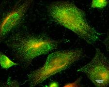
ULK1 Antibody - Immunofluorescence analysis.
Select
ULK1 Rabbit anti-Human Polyclonal (aa400-499) Antibody
Human
IF, WB
Unconjugated
20 µl/$275; 50 µl/$311; 100 µl/$389
ULK1 Antibody - Immunocytochemistry/Immunofluorescence analysis using Rabbit Anti-ULK1 Polyclonal Antibody. Tissue: Neuroblastoma cell line (SK-N-BE). Species: Human. Fixation: 4% Formaldehyde for 15 min at RT. Primary Antibody: Rabbit Anti-ULK1 Polyclonal Antibody  at 1:100 for 60 min at RT. Secondary Antibody: Goat Anti-Rabbit ATTO 488 at 1:200 for 60 min at RT. Counterstain: Phalloidin Texas Red F-Actin stain; DAPI (blue) nuclear stain at 1:1000, 1:5000 for 60 min at RT, 5 min at RT. Localization: Cytoplasm, Preautophagosomal Structure. Magnification: 60X. (A) DAPI (blue) nuclear stain (B) Phalloidin Texas Red F-Actin stain (C) ULK1 Antibody (D) Composite.
Select
ULK1 Rabbit anti-Human Polyclonal (Biotin) Antibody
Mouse, Rat, Human
IF, WB
Biotin Conjugated
100 µg/$477
ULK1 Antibody - Immunocytochemistry/Immunofluorescence analysis using Rabbit Anti-ULK1 Polyclonal Antibody. Tissue: Neuroblastoma cell line (SK-N-BE). Species: Human. Fixation: 4% Formaldehyde for 15 min at RT. Primary Antibody: Rabbit Anti-ULK1 Polyclonal Antibody  at 1:100 for 60 min at RT. Secondary Antibody: Goat Anti-Rabbit ATTO 488 at 1:200 for 60 min at RT. Counterstain: Phalloidin Texas Red F-Actin stain; DAPI (blue) nuclear stain at 1:1000, 1:5000 for 60 min at RT, 5 min at RT. Localization: Cytoplasm, Preautophagosomal Structure. Magnification: 60X. (A) DAPI (blue) nuclear stain (B) Phalloidin Texas Red F-Actin stain (C) ULK1 Antibody (D) Composite.
Select
ULK1 Rabbit anti-Human Polyclonal (FITC) Antibody
Mouse, Rat, Human
IF, WB
FITC Conjugated
100 µg/$473
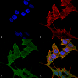
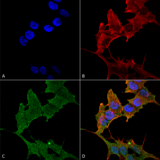
ULK1 Antibody - Immunocytochemistry/Immunofluorescence analysis using Rabbit Anti-ULK1 Polyclonal Antibody. Tissue: Neuroblastoma cell line (SK-N-BE). Species: Human. Fixation: 4% Formaldehyde for 15 min at RT. Primary Antibody: Rabbit Anti-ULK1 Polyclonal Antibody  at 1:100 for 60 min at RT. Secondary Antibody: Goat Anti-Rabbit ATTO 488 at 1:200 for 60 min at RT. Counterstain: Phalloidin Texas Red F-Actin stain; DAPI (blue) nuclear stain at 1:1000, 1:5000 for 60 min at RT, 5 min at RT. Localization: Cytoplasm, Preautophagosomal Structure. Magnification: 60X. (A) DAPI (blue) nuclear stain (B) Phalloidin Texas Red F-Actin stain (C) ULK1 Antibody (D) Composite.
Select
ULK1 Rabbit anti-Human Polyclonal (RPE) Antibody
Mouse, Rat, Human
IF, WB
RPE Conjugated
100 µg/$477
ULK1 Antibody - Immunocytochemistry/Immunofluorescence analysis using Rabbit Anti-ULK1 Polyclonal Antibody. Tissue: Neuroblastoma cell line (SK-N-BE). Species: Human. Fixation: 4% Formaldehyde for 15 min at RT. Primary Antibody: Rabbit Anti-ULK1 Polyclonal Antibody  at 1:100 for 60 min at RT. Secondary Antibody: Goat Anti-Rabbit ATTO 488 at 1:200 for 60 min at RT. Counterstain: Phalloidin Texas Red F-Actin stain; DAPI (blue) nuclear stain at 1:1000, 1:5000 for 60 min at RT, 5 min at RT. Localization: Cytoplasm, Preautophagosomal Structure. Magnification: 60X. (A) DAPI (blue) nuclear stain (B) Phalloidin Texas Red F-Actin stain (C) ULK1 Antibody (D) Composite.
Select
ULK1 Rabbit anti-Human Polyclonal (Atto 390) Antibody
Mouse, Rat, Human
IF, WB
Atto 390 Conjugated
100 µg/$480
ULK1 Antibody - Immunocytochemistry/Immunofluorescence analysis using Rabbit Anti-ULK1 Polyclonal Antibody. Tissue: Neuroblastoma cell line (SK-N-BE). Species: Human. Fixation: 4% Formaldehyde for 15 min at RT. Primary Antibody: Rabbit Anti-ULK1 Polyclonal Antibody  at 1:100 for 60 min at RT. Secondary Antibody: Goat Anti-Rabbit ATTO 488 at 1:200 for 60 min at RT. Counterstain: Phalloidin Texas Red F-Actin stain; DAPI (blue) nuclear stain at 1:1000, 1:5000 for 60 min at RT, 5 min at RT. Localization: Cytoplasm, Preautophagosomal Structure. Magnification: 60X. (A) DAPI (blue) nuclear stain (B) Phalloidin Texas Red F-Actin stain (C) ULK1 Antibody (D) Composite.
Select
ULK1 Rabbit anti-Human Polyclonal (PerCP) Antibody
Mouse, Rat, Human
IF, WB
PerCP Conjugated
100 µg/$479
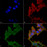
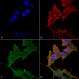
ULK1 Antibody - Immunocytochemistry/Immunofluorescence analysis using Rabbit Anti-ULK1 Polyclonal Antibody. Tissue: Neuroblastoma cell line (SK-N-BE). Species: Human. Fixation: 4% Formaldehyde for 15 min at RT. Primary Antibody: Rabbit Anti-ULK1 Polyclonal Antibody  at 1:100 for 60 min at RT. Secondary Antibody: Goat Anti-Rabbit ATTO 488 at 1:200 for 60 min at RT. Counterstain: Phalloidin Texas Red F-Actin stain; DAPI (blue) nuclear stain at 1:1000, 1:5000 for 60 min at RT, 5 min at RT. Localization: Cytoplasm, Preautophagosomal Structure. Magnification: 60X. (A) DAPI (blue) nuclear stain (B) Phalloidin Texas Red F-Actin stain (C) ULK1 Antibody (D) Composite.
Select
ULK1 Rabbit anti-Human Polyclonal (Atto 594) Antibody
Mouse, Rat, Human
IF, WB
Atto 594 Conjugated
100 µg/$479
ULK1 Antibody - Immunocytochemistry/Immunofluorescence analysis using Rabbit Anti-ULK1 Polyclonal Antibody. Tissue: Neuroblastoma cell line (SK-N-BE). Species: Human. Fixation: 4% Formaldehyde for 15 min at RT. Primary Antibody: Rabbit Anti-ULK1 Polyclonal Antibody  at 1:100 for 60 min at RT. Secondary Antibody: Goat Anti-Rabbit ATTO 488 at 1:200 for 60 min at RT. Counterstain: Phalloidin Texas Red F-Actin stain; DAPI (blue) nuclear stain at 1:1000, 1:5000 for 60 min at RT, 5 min at RT. Localization: Cytoplasm, Preautophagosomal Structure. Magnification: 60X. (A) DAPI (blue) nuclear stain (B) Phalloidin Texas Red F-Actin stain (C) ULK1 Antibody (D) Composite.
Select
ULK1 Rabbit anti-Human Polyclonal (Atto 488) Antibody
Mouse, Rat, Human
IF, WB
Atto 488 Conjugated
100 µg/$479
ULK1 Antibody - Immunocytochemistry/Immunofluorescence analysis using Rabbit Anti-ULK1 Polyclonal Antibody. Tissue: Neuroblastoma cell line (SK-N-BE). Species: Human. Fixation: 4% Formaldehyde for 15 min at RT. Primary Antibody: Rabbit Anti-ULK1 Polyclonal Antibody  at 1:100 for 60 min at RT. Secondary Antibody: Goat Anti-Rabbit ATTO 488 at 1:200 for 60 min at RT. Counterstain: Phalloidin Texas Red F-Actin stain; DAPI (blue) nuclear stain at 1:1000, 1:5000 for 60 min at RT, 5 min at RT. Localization: Cytoplasm, Preautophagosomal Structure. Magnification: 60X. (A) DAPI (blue) nuclear stain (B) Phalloidin Texas Red F-Actin stain (C) ULK1 Antibody (D) Composite.
Select
ULK1 Rabbit anti-Human Polyclonal (APC) Antibody
Mouse, Rat, Human
IF, WB
APC Conjugated
100 µg/$479
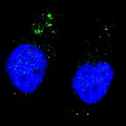
ULK1 Antibody - Fluorescent image of U251 cells stained with ULK1 (phospho S556) antibody. U251 cells were treated with Chloroquine (50 mu M,16h), then fixed with 4% PFA (20 min), permeabilized with Triton X-100 (0.2%, 30 min). Cells were then incubated ULK1 (phospho S556) primary antibody (1:200, 2 h at room temperature). For secondary antibody, Alexa Fluor 488 conjugated donkey anti-rabbit antibody (green) was used (1:1000, 1h). Nuclei were counterstained with Hoechst 33342 (blue) (10 ug/ml, 5 min). ULK1 (phospho S556) immunoreactivity is localized to autophagic vacuoles in the cytoplasm of U251 cells.
Select
ULK1 Rabbit anti-Human Polyclonal (pSer556) Antibody
Human
DB, IF
Unconjugated
400 µl/$393
Viewing 1-25 of 89 product results


Filtered By:
Products: Antibodies


If you do not find the reagent or information you require, please contact Customer.Support@LSBio.com to inquire about additional products in development.

PLEASE NOTE

For RESEARCH USE ONLY. Intended for use by laboratory professionals. Not intended for human diagnostic or therapeutic purposes.

The data on this page has been compiled from LifeSpan internal sources, the National Center for Biotechnology Information (NCBI), and The Universal Protein Resource (UniProt).