Products
Research Areas
COVID-19
Resources
Login
Quick Order
Cart Cart lightblue
Login
Registration enables users to use special features of this website, such as past
order histories, retained contact details for faster checkout, review submissions, and special promotions.


Fields marked with a * are required.

Login
Quick Order
Contact Us

Location


Corporate Headquarters

Vector Laboratories, Inc.
6737 Mowry Ave
Newark, CA 94560
United States

Telephone Numbers


Customer Service: (800) 227-6666 / (650) 697-3600


Contact Us


Additional Contact Details

Login
Registration enables users to use special features of this website, such as past
order histories, retained contact details for faster checkout, review submissions, and special promotions.


Fields marked with a * are required.

Login
Quick Order

PRKD1 / PKC Mu

Protein kinase D1

Serine/threonine-protein kinase that converts transient diacylglycerol (DAG) signals into prolonged physiological effects downstream of PKC, and is involved in the regulation of MAPK8/JNK1 and Ras signaling, Golgi membrane integrity and trafficking, cell survival through NF-kappa-B activation, cell migration, cell differentiation by mediating HDAC7 nuclear export, cell proliferation via MAPK1/3 (ERK1/2) signaling, and plays a role in cardiac hypertrophy, VEGFA-induced angiogenesis, genotoxic-induced apoptosis and flagellin-stimulated inflammatory response. Phosphorylates the epidermal growth factor receptor (EGFR) on dual threonine residues, which leads to the suppression of epidermal growth factor (EGF)-induced MAPK8/JNK1 activation and subsequent JUN phosphorylation. Phosphorylates RIN1, inducing RIN1 binding to 14-3-3 proteins YWHAB, YWHAE and YWHAZ and increased competition with RAF1 for binding to GTP-bound form of Ras proteins (NRAS, HRAS and KRAS). Acts downstream of the heterotrimeric G-protein beta/gamma-subunit complex to maintain the structural integrity of the Golgi membranes, and is required for protein transport along the secretory pathway. In the trans-Golgi network (TGN), regulates the fission of transport vesicles that are on their way to the plasma membrane. May act by activating the lipid kinase phosphatidylinositol 4-kinase beta (PI4KB) at the TGN for the local synthesis of phosphorylated inositol lipids, which induces a sequential production of DAG, phosphatidic acid (PA) and lyso-PA (LPA) that are necessary for membrane fission and generation of specific transport carriers to the cell surface. Under oxidative stress, is phosphorylated at Tyr-463 via SRC-ABL1 and contributes to cell survival by activating IKK complex and subsequent nuclear translocation and activation of NFKB1. Involved in cell migration by regulating integrin alpha-5/beta-3 recycling and promoting its recruitment in newly forming focal adhesion. In osteoblast differentiation, mediates the bone morphogenic protein 2 (BMP2)-induced nuclear export of HDAC7, which results in the inhibition of HDAC7 transcriptional repression of RUNX2. In neurons, plays an important role in neuronal polarity by regulating the biogenesis of TGN-derived dendritic vesicles, and is involved in the maintenance of dendritic arborization and Golgi structure in hippocampal cells. May potentiate mitogenesis induced by the neuropeptide bombesin or vasopressin by mediating an increase in the duration of MAPK1/3 (ERK1/2) signaling, which leads to accumulation of immediate-early gene products including FOS that stimulate cell cycle progression. Plays an important role in the proliferative response induced by low calcium in keratinocytes, through sustained activation of MAPK1/3 (ERK1/2) pathway. Downstream of novel PKC signaling, plays a role in cardiac hypertrophy by phosphorylating HDAC5, which in turn triggers XPO1/CRM1-dependent nuclear export of HDAC5, MEF2A transcriptional activation and induction of downstream target genes that promote myocyte hypertrophy and pathological cardiac remodeling. Mediates cardiac troponin I (TNNI3) phosphorylation at the PKA sites, which results in reduced myofilament calcium sensitivity, and accelerated crossbridge cycling kinetics. The PRKD1-HDAC5 pathway is also involved in angiogenesis by mediating VEGFA-induced specific subset of gene expression, cell migration, and tube formation. In response to VEGFA, is necessary and required for HDAC7 phosphorylation which induces HDAC7 nuclear export and endothelial cell proliferation and migration. During apoptosis induced by cytarabine and other genotoxic agents, PRKD1 is cleaved by caspase-3 at Asp-378, resulting in activation of its kinase function and increased sensitivity of cells to the cytotoxic effects of genotoxic agents. In epithelial cells, is required for transducing flagellin-stimulated inflammatory responses by binding and phosphorylating TLR5, which contributes to MAPK14/p38 activation and production of inflammatory cytokines. May play a role in inflammatory response by mediating activation of NF-kappa-B. May be involved in pain transmission by directly modulating TRPV1 receptor.

Gene Name: Protein kinase D1
Family/Subfamily: Protein Kinase , PKD
Synonyms: PRKD1, PKCM, Pkcmu, Protein kinase C, mu, Protein kinase D, NPKC-mu, PRKCM, Protein kinase C mu, Protein kinase D1, NPKC-D1, PKC-MU, PKD, Protein kinase C mu type
Target Sequences: NM_002742 CAA53384.1 Q15139

☰ Filters
Products
Antibodies (75)
Primary (75)
PRKD1 / PKC Mu (75)
Human (67)
Mouse (63)
Rat (44)
Bovine (4)
Chicken (2)
Dog (6)
Horse (4)
Monkey (6)
Pig (4)
Rabbit (4)
Zebrafish (2)
IHC (35)
IHC-P (10)
WB (72)
ELISA (25)
ICC (1)
IF (9)
IP (3)
Peptide-ELISA (17)
rabbit (73)
goat (2)
Product Group
IHCPlus (1)
IgG (45)
polyclonal pc (75)
APC (2)
APC Conjugated (3)
Azide-free (1)
Biotin Conjugated (3)
Cy3 Conjugated (2)
Cy7 Conjugated (2)
FITC Conjugated (3)
HRP Conjugated (3)
PE Conjugated (3)
Unconjugated (56)
aa349-612 (8)
aa651-876 (8)
aa361-389 (7)
pSer910 (6)
pTyr463 (4)
140-220 aa (2)
pSer205 (2)
pSer738 (2)
400-480 aa (1)
850-930 aa (1)
Arg201 (1)
C-Terminus (1)
Gly459 (1)
Ile740 (1)
Internal (1)
aa171-220 (1)
aa233-246 (1)
aa383-395 (1)
aa429-478 (1)
aa529-862 (1)
aa695-744 (1)
aa706-755 (1)
aa863-912 (1)
p205 (1)
p463 (1)
p744/748 (1)
p910 (1)
pSer738+Ser742 (1)
pSer738,pSer742 (1)
pSer738/Ser742 (1)
pSer744/748 (1)
pSer744/Ser748 (1)
pSer916 (1)
No (74)
Yes (1)
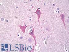
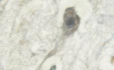
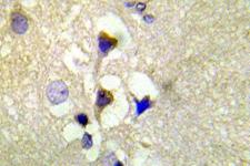
PRKD1 / PKC Mu Antibody - Anti-PKC Mu antibody IHC of human brain, cortex neurons. Immunohistochemistry of formalin-fixed, paraffin-embedded tissue after heat-induced antigen retrieval. Antibody dilution 1:67.
Select
PRKD1 / PKC Mu Rabbit anti-Human Polyclonal (aa171-220) Antibody
Mouse, Rat, Human
IF, IHC, IHC-P, Peptide-ELISA, WB
Unconjugated
50 µl/$460
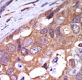
PRKD1 / PKC Mu Antibody - Formalin-fixed and paraffin-embedded human cancer tissue reacted with the primary antibody, which was peroxidase-conjugated to the secondary antibody, followed by AEC staining. This data demonstrates the use of this antibody for immunohistochemistry; clinical relevance has not been evaluated. BC = breast carcinoma; HC = hepatocarcinoma.
Select
PRKD1 / PKC Mu Rabbit anti-Human Polyclonal (aa361-389) Antibody
Mouse, Human
IHC, IHC-P, WB
Unconjugated
400 µl/$393
PRKD1 / PKC Mu Antibody - HEK293 lysate overexpressing Human DYKDDDDK-tagged PKD1 was used to immunoprecipitate PKD1 with 2ug antibody. The precipitate was subsequently probed in Western blot using antibody at 1ug/ml. The secondary anti-goat picks up the heavy chain of antibody used
Select
PRKD1 / PKC Mu Goat anti-Human Polyclonal (aa233-246) Antibody
Human
IHC, IP, Peptide-ELISA, WB
Unconjugated
100 µg/$503
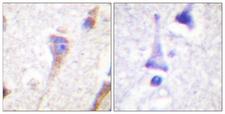
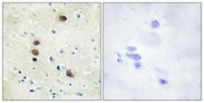
PRKD1 / PKC Mu Antibody - Immunohistochemistry analysis of paraffin-embedded human brain tissue, using PKD1/PKC mu Antibody. The picture on the right is blocked with the synthesized peptide.
Select
PRKD1 / PKC Mu Rabbit anti-Human Polyclonal (aa429-478) Antibody
Mouse, Rat, Human
IHC, IHC-P, Peptide-ELISA, WB
Unconjugated
50 µl/$334; 100 µl/$397
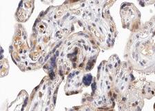
PRKD1 / PKC Mu Antibody - 1:100 staining human Placenta tissue by IHC-P. The tissue was formaldehyde fixed and a heat mediated antigen retrieval step in citrate buffer was performed. The tissue was then blocked and incubated with the antibody for 1.5 hours at 22°C. An HRP conjugated goat anti-rabbit antibody was used as the secondary.
Select
PRKD1 / PKC Mu Rabbit anti-Human Polyclonal (pSer738,pSer742) Antibody
Mouse, Rat, Human
IF, IHC, Peptide-ELISA, WB
Unconjugated
100 µl/$379; 200 µl/$421
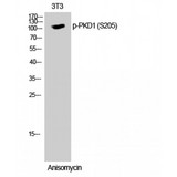
PRKD1 / PKC Mu Antibody - Western blot of Phospho-PKD1 (S205) antibody
Select
PRKD1 / PKC Mu Rabbit anti-Human Polyclonal (140-220 aa) Antibody
Mouse, Rat, Human
ELISA, IHC, WB
Unconjugated
50 µg/$295; 100 µg/$335; 200 µg/$394
Select
PRKD1 / PKC Mu Rabbit anti-Mouse Polyclonal (pSer744/Ser748) Antibody
Mouse, Rat, Human, Monkey
ELISA, WB
Unconjugated
100 µl/$717
PRKD1 / PKC Mu Antibody - Peptide - + Immunohistochemistry analysis of paraffin-embedded human brain tissue using PKD1/PKC µ (Ab-205) antibody.
Select
PRKD1 / PKC Mu Rabbit anti-Human Polyclonal (p205) Antibody
Mouse, Human
ELISA, IF, IHC, WB
Unconjugated
100 µg/$357
PRKD1 / PKC Mu Antibody - 1:200 staining CRC tissue by IHC-P.
Select
PRKD1 / PKC Mu Rabbit anti-Human Polyclonal (pTyr463) Antibody
Mouse, Rat, Human
IF, IHC, Peptide-ELISA, WB
Unconjugated
100 µl/$379; 200 µl/$421
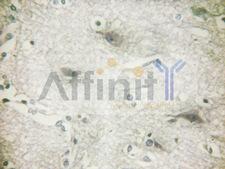
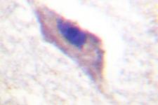
PRKD1 / PKC Mu Antibody - 1:200 staining human brain tissue by IHC-P. The tissue was formaldehyde fixed and a heat mediated antigen retrieval step in citrate buffer was performed. The tissue was then blocked and incubated with the antibody for 1.5 hours at 22°C. An HRP conjugated goat anti-rabbit antibody was used as the secondary.
Select
PRKD1 / PKC Mu Rabbit anti-Human Polyclonal Antibody
Mouse, Rat, Human
IF, IHC, Peptide-ELISA, WB
Unconjugated
100 µl/$379; 200 µl/$421
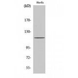
PRKD1 / PKC Mu Antibody - Western blot of PKD1 antibody
Select
PRKD1 / PKC Mu Rabbit anti-Human Polyclonal (140-220 aa) Antibody
Mouse, Rat, Human
ELISA, IF, IHC, WB
Unconjugated
50 µg/$295; 100 µg/$335; 200 µg/$394
PRKD1 / PKC Mu Antibody - Immunohistochemistry analysis of phospho-PKD1/PKC-mu (Phospho-Ser910) antibody in paraffin-embedded human brain tissue.
Select
PRKD1 / PKC Mu Rabbit anti-Human Polyclonal (pSer910) Antibody
Mouse, Rat, Human
ELISA, IHC, Peptide-ELISA, WB
Unconjugated
50 µl/$334; 100 µl/$397
PRKD1 / PKC Mu Antibody - IHC of PKD1/PKC (R201) pAb in paraffin-embedded human breast tissue.
Select
PRKD1 / PKC Mu Rabbit anti-Human Polyclonal (Arg201) Antibody
Mouse, Rat, Human
IF, IHC, IHC-P, WB
Unconjugated
50 µl/$321; 100 µl/$403
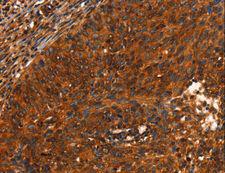
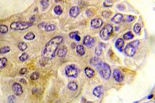
PRKD1 / PKC Mu Antibody - Immunohistochemistry of paraffin-embedded Human cervical cancer using PRKD1 Polyclonal Antibody at dilution of 1:40.
Select
PRKD1 / PKC Mu Rabbit anti-Human Polyclonal Antibody
Mouse, Rat, Human
ELISA, IHC, WB
Unconjugated
20 µl/$254; 60 µl/$296; 120 µl/$355; 200 µl/$450
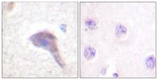
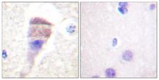
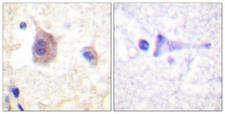
PRKD1 / PKC Mu Antibody - Immunohistochemistry analysis of paraffin-embedded human brain, using PKD1/PKC mu (Phospho-Ser205) Antibody. The picture on the right is blocked with the phospho peptide.
Select
PRKD1 / PKC Mu Rabbit anti-Human Polyclonal (pSer205) Antibody
Mouse, Rat, Human
IHC, IHC-P, Peptide-ELISA, WB
Unconjugated
50 µl/$334; 100 µl/$397
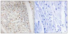
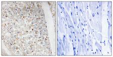
PRKD1 / PKC Mu Antibody - Immunohistochemistry analysis of paraffin-embedded human heart, using PKD1/2/3/PKC mu (Phospho-Ser738+Ser742) Antibody. The picture on the right is blocked with the phospho peptide.
Select
PRKD1 / PKC Mu Rabbit anti-Human Polyclonal (pSer738/Ser742) Antibody
Mouse, Rat, Human
IHC, IHC-P, Peptide-ELISA, WB
Unconjugated
50 µl/$334; 100 µl/$397
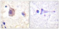
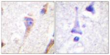
PRKD1 / PKC Mu Antibody - Immunohistochemistry analysis of paraffin-embedded human brain, using PKD1/PKC mu (Phospho-Tyr463) Antibody. The picture on the right is blocked with the phospho peptide.
Select
PRKD1 / PKC Mu Rabbit anti-Human Polyclonal (pTyr463) Antibody
Mouse, Rat, Human
IHC, IHC-P, Peptide-ELISA, WB
Unconjugated
50 µl/$334; 100 µl/$397
Select
PRKD1 / PKC Mu Rabbit anti-Human Polyclonal (aa361-389) Antibody
Mouse, Human
ELISA, IHC, WB
Unconjugated
200 µl/$734
Select
PRKD1 / PKC Mu Rabbit anti-Human Polyclonal (400-480 aa) Antibody
Mouse, Rat, Human
ELISA, IHC, WB
Unconjugated
50 µg/$295; 100 µg/$335; 200 µg/$394
PRKD1 / PKC Mu Antibody - P-peptide - + Immunohistochemistry analysis of paraffin-embedded human brain tissue using PKD1/PKC µ (Phospho-Ser205) antibody.
Select
PRKD1 / PKC Mu Rabbit anti-Human Polyclonal (pSer205) Antibody
Mouse, Human
ELISA, IHC, WB
Unconjugated
100 µg/$357
PRKD1 / PKC Mu Antibody - P-peptide - + Immunohistochemistry analysis of paraffin-embedded human brain tissue using PKD1/PKC µ (Phospho-Tyr463) antibody.
Select
PRKD1 / PKC Mu Rabbit anti-Human Polyclonal (pTyr463) Antibody
Mouse, Rat, Human
ELISA, IHC, WB
Unconjugated
100 µg/$357
PRKD1 / PKC Mu Antibody - Peptide - + Immunohistochemistry analysis of paraffin-embedded human brain tissue using PKD1/PKC µ (Ab-463) antibody.
Select
PRKD1 / PKC Mu Rabbit anti-Human Polyclonal (p463) Antibody
Mouse, Rat, Human
ELISA, IHC, WB
Unconjugated
100 µg/$357
PRKD1 / PKC Mu Antibody - P-peptide - + Immunohistochemistry analysis of paraffin-embedded human heart tissue using PKD1/2/3/PKC µ (Phospho-Ser738+Ser742) antibody.
Select
PRKD1 / PKC Mu Rabbit anti-Human Polyclonal (pSer738+Ser742) Antibody
Mouse, Rat, Human
ELISA, IHC, WB
Unconjugated
100 µg/$357
PRKD1 / PKC Mu Antibody - Immunohistochemistry analysis of phospho-PKD1/PKC-mu (Phospho-Ser738) antibody in paraffin-embedded human brain tissue.
Select
PRKD1 / PKC Mu Rabbit anti-Human Polyclonal (pSer738) Antibody
Mouse, Rat, Human
ELISA, IHC, Peptide-ELISA
Unconjugated
50 µl/$334; 100 µl/$397
PRKD1 / PKC Mu Antibody - Staining A549 cells by IF/ICC. The samples were fixed with PFA and permeabilized in 0.1% Triton X-100, then blocked in 10% serum for 45 min at 25°C. The primary antibody was diluted at 1:200 and incubated with the sample for 1 hour at 37°C. An Alexa Fluor 594 conjugated goat anti-rabbit IgG (H+L) antibody, diluted at 1/600, was used as secondary antibody.
Select
PRKD1 / PKC Mu Rabbit anti-Human Polyclonal Antibody
Mouse, Rat, Human
IF, Peptide-ELISA, WB
Unconjugated
100 µl/$379; 200 µl/$421
Viewing 1-25 of 75 product results


Filtered By:
Products: Antibodies


If you do not find the reagent or information you require, please contact Customer.Support@LSBio.com to inquire about additional products in development.

PLEASE NOTE

For RESEARCH USE ONLY. Intended for use by laboratory professionals. Not intended for human diagnostic or therapeutic purposes.

The data on this page has been compiled from LifeSpan internal sources, the National Center for Biotechnology Information (NCBI), and The Universal Protein Resource (UniProt).