Products
Research Areas
COVID-19
Resources
Login
Quick Order
Cart Cart lightblue
Login
Registration enables users to use special features of this website, such as past
order histories, retained contact details for faster checkout, review submissions, and special promotions.


Fields marked with a * are required.

Login
Quick Order
Contact Us

Location


Corporate Headquarters

Vector Laboratories, Inc.
6737 Mowry Ave
Newark, CA 94560
United States

Telephone Numbers


Customer Service: (800) 227-6666 / (650) 697-3600


Contact Us


Additional Contact Details

Login
Registration enables users to use special features of this website, such as past
order histories, retained contact details for faster checkout, review submissions, and special promotions.


Fields marked with a * are required.

Login
Quick Order

CD274 / B7-H1 / PD-L1

CD274 molecule

CD274, also known as B7-H1 and PD-L1, a cell surface glycoprotein, is a member of the B7 family of co-stimulatory molecules. CD274 is expressed constitutively on macrophages and dendritic cells, and is induced on activated T-cells, B-cells, endothelial cells and epithelial cells in response to interferons. CD274 has dual functions: inhibition of activated effector T cells and co-stimulation of naïve T cells. It inhibits proliferation of activated T cells via ligation to the co-inhibitory molecule CD279 (programmed death-1; PD-1) leading to the secretion of the regulatory cytokine interleukin-10. Expression of CD274 in tumor cells is considered to be prognostic in many types of human malignancies, including colon cancer and renal cell carcinoma.

Gene Name: CD274 molecule
Synonyms: CD274, B7-H, B7 homolog 1, B7-H1, B7H1, CD274 molecule, CD274 antigen, PDCD1 ligand 1, PDL1, PD-L1, PDCD1L1, PDCD1LG1, Programmed death ligand 1
Target Sequences: NM_014143 NP_054862.1 Q9NZQ7

Publications (19)

1
BRAF inhibition is associated with enhanced melanoma antigen expression and a more favorable tumor microenvironment in patients with metastatic melanoma. Frederick DT, Piris A, Cogdill AP, Cooper ZA, Lezcano C, Ferrone CR, Mitra D, Boni A, Newton LP, Liu C, Peng W, Sullivan RJ, Lawrence DP, Hodi FS, Overwijk WW, Lizée G, Murphy GF, Hwu P, Flaherty KT, Fisher DE, Wargo JA. Clinical cancer research : an official journal of the American Association for Cancer Research. 2013 19:1225-31. (WB; Human) [Full Text Article] [PubMed:23307859] [PMC:PMC3752683]
2
Programmed cell death protein 1 and programmed death-ligand 1 are expressed on the surface of some small-cell lung cancer lines. Yamane H, Isozaki H, Takeyama M, Ochi N, Kudo K, Honda Y, Yamagishi T, Kubo T, Kiura K, Takigawa N. American journal of cancer research. 2015 5:1553-7. (Flo; Human) [PubMed:26101718] [PMC:PMC4473331]
3
Characterization of myeloid leukocytes and soluble mediators in pancreatic cancer: importance of myeloid-derived suppressor cells. Karakhanova S, Link J, Heinrich M, Shevchenko I, Yang Y, Hassenpflug M, Bunge H, von Ahn K, Brecht R, Mathes A, Maier C, Umansky V, Werner J, Bazhin AV. Oncoimmunology. 2015 4:e998519. (Flo; Mouse) [Full Text Article] [PubMed:26137414] [PMC:PMC4485765]
4
PD-1 mediates functional exhaustion of activated NK cells in patients with Kaposi sarcoma. Beldi-Ferchiou A, Lambert M, Dogniaux S, Vély F, Vivier E, Olive D, Dupuy S, Levasseur F, Zucman D, Lebbé C, Sène D, Hivroz C, Caillat-Zucman S. Oncotarget. 2016 7:72961-72977. (WB; Human) [Full Text Article] [PubMed:27662664] [PMC:PMC5341956]
5
EML4-ALK enhances programmed cell death-ligand 1 expression in pulmonary adenocarcinoma via hypoxia-inducible factor (HIF)-1a and STAT3. Koh J, Jang JY, Keam B, Kim S, Kim MY, Go H, Kim TM, Kim DW, Kim CW, Jeon YK, Chung DH. Oncoimmunology. 2016 5:e1108514. (Flo; Human) [Full Text Article] [PubMed:27141364]
6
Induction of PD-L1 Expression by the EML4-ALK Oncoprotein and Downstream Signaling Pathways in Non-Small Cell Lung Cancer. Ota K, Azuma K, Kawahara A, Hattori S, Iwama E, Tanizaki J, Harada T, Matsumoto K, Takayama K, Takamori S, Kage M, Hoshino T, Nakanishi Y, Okamoto I. Clinical cancer research : an official journal of the American Association for Cancer Research. 2015 21:4014-21. (WB; Human) [Full Text Article] [PubMed:26019170]
7
Association of PD-L1 overexpression with activating EGFR mutations in surgically resected nonsmall-cell lung cancer. Azuma K, Ota K, Kawahara A, Hattori S, Iwama E, Harada T, Matsumoto K, Takayama K, Takamori S, Kage M, Hoshino T, Nakanishi Y, Okamoto I. Annals of oncology : official journal of the European Society for Medical Oncology / ESMO. 2014 25:1935-40. (WB; Human) [Full Text Article] [PubMed:25009014]
8
Neuronal programmed cell death-1 ligand expression regulates retinal ganglion cell number in neonatal and adult mice. Sham CW, Chan AM, Kwong JM, Caprioli J, Nusinowitz S, Chen B, Lee JG, Gandhi NM, Francisco LM, Sharpe AH, Chen L, Braun J, Gordon LK. Journal of neuro-ophthalmology : the official journal of the North American Neuro-Ophthalmology Society. 2012 32:227-37. (WB; Human) [Full Text Article] [PubMed:22635166]
9
Clinicopathological and prognostic significance of programmed cell death ligand 1 (PD-L1) expression in patients with esophageal squamous cell carcinoma: a meta-analysis. Qu HX, Zhao LP, Zhan SH, Geng CX, Xu L, Xin YN, Jiang XJ. Journal of thoracic disease. 2016 November;8:3197-3204. (Human) [Full Text Article] [PubMed:28066599] [PMC:PMC5179395]
10
High programmed cell death 1 ligand-1 expression: association with CD8+ T-cell infiltration and poor prognosis in human medulloblastoma. Murata D, Mineharu Y, Arakawa Y, Liu B, Tanji M, Yamaguchi M, Fujimoto KI, Fukui N, Terada Y, Yokogawa R, Yamaguchi M, Minamiguchi S, Miyamoto S. Journal of neurosurgery. 2018 March;128:710-716. [Full Text Article] [PubMed:28474991]
11
Prognostic significance of programmed death ligand-1 immunohistochemical expression in esophageal cancer: A meta-analysis of the literature. Yu W, Guo Y. Medicine. 2018 July;97:e11614. [Full Text Article] [PubMed:30045299] [PMC:PMC6078683]
12
Oncogenic kinase NPM/ALK induces through STAT3 expression of immunosuppressive protein CD274 (PD-L1, B7-H1). Michal Marzec, Qian Zhang, Ami Goradia, Puthiyaveettil N Raghunath, Xiaobin Liu, Michele Paessler, Hong Yi Wang, Maria Wysocka, Mangeng Cheng, Bruce A Ruggeri, Mariusz A Wasik. Proceedings of the National Academy of Sciences of the United States of America. 2008 December;105:20852-7. [Full Text Article] [PubMed:19088198] [PMC:PMC2634900]
13
Juxtacrine Signaling Inhibits Antitumor Immunity by Upregulating PD-L1 Expression. Wen-Hao Yang, Jong-Ho Cha, Weiya Xia, Heng-Huan Lee, Li-Chuan Chan, Ying-Nai Wang, Jennifer L Hsu, Guoxin Ren, Mien-Chie Hung. Cancer research. 2018 July;78:3761-3768. [Full Text Article] [PubMed:29789418] [PMC:PMC6050079] Related Antibodies: LS-C338364.
14
PD-L1-mediated gasdermin C expression switches apoptosis to pyroptosis in cancer cells and facilitates tumour necrosis. Junwei Hou, Rongce Zhao, Weiya Xia, Chiung-Wen Chang, Yun You, Jung-Mao Hsu, Lei Nie, Yeh Chen, Yu-Chuan Wang, Chunxiao Liu, Wei-Jan Wang, Yun Wu, Baozhen Ke, Jennifer L Hsu, Kebin Huang, Zu Ye, Yi Yang, Xianghou Xia, Yintao Li, Chia-Wei Li, Bin Shao, John A Tainer, Mien-Chie Hung. Nature cell biology. 2020 October;22:1264-1275. [Full Text Article] [PubMed:32929201] [PMC:PMC7653546] Related Antibodies: LS-C338364.
15
anlotinib alters tumor immune microenvironment by downregulating PD-L1 expression on vascular endothelial cells. Shaochuan Liu , Tingting Qin , Zhujun Liu, Jing Wang, Yanan Jia, Yingfang Feng, Yuan Gao, Kai Li. Cell death & disease. 2020 May;11:309. [Full Text Article] [PubMed:32366856] [PMC:PMC7198575] Related Antibodies: LS-C746930.
16
Tumor Lymphatic Function Regulates Tumor Inflammatory and Immunosuppressive Microenvironments. Raghu P Kataru, Catherine L Ly, Jinyeon Shin, Hyeung Ju Park, Jung Eun Baik, Sonia Rehal, Sagrario Ortega, David Lyden, Babak J Mehrara. Cancer immunology research. 2019 Aug;7:1345-1358. [Full Text Article] [PubMed:31186247] [PMC:PMC6677612]
17
Metformin Promotes Antitumor Immunity via Endoplasmic-Reticulum-Associated Degradation of PD-L110<sup> Department of Experimental Radiation Oncology, University of Texas MD Anderson Cancer Center, Houston, TX 77030, USA12<sup> Department of Breast Surgical Oncology, University of Texas MD Anderson Cancer Center, Houston, TX 77030, USA14<sup> Department of Experimental Radiation Oncology, University of Texas MD Anderson Cancer Center, Houston, TX 77030, USA; Graduate School of Biomedical Sciences. Jong-Ho Cha, Wen-Hao Yang, Weiya Xia, Yongkun Wei, Li-Chuan Chan, Seung-Oe Lim, Chia-Wei Li, Taewan Kim, Shih-Shin Chang, Heng-Huan Lee, Jennifer L Hsu, Hung-Ling Wang, Chu-Wei Kuo, Wei-Chao Chang, Sirwan Hadad, Colin A Purdie, Aaron M McCoy, Shirong Cai, Yizheng Tu, Jennifer K Litton, Elizabeth A Mittendorf, Stacy L Moulder, William F Symmans, Alastair M Thompson, Helen Piwnica-Worms, Chung-Hsuan Chen, Kay-Hooi Khoo, Mien-Chie Hung. Molecular cell. 2018 Aug;71:606-620.e7. [Full Text Article] [PubMed:30118680] [PMC:PMC6786495] Related Antibodies: LS-C338364.
18
A meta-analysis of CD274 (PD-L1) assessment and prognosis in colorectal cancer and its role in predicting response to anti-PD-1 therapy. Peter G Alexander, Donald C McMillan, James H Park. Critical reviews in oncology/hematology. 2020 November;157:103147. [Full Text Article] [PubMed:33278675] Related Antibodies: LS-C179482.
19
The CD155/TIGIT axis promotes and maintains immune evasion in neoantigen-expressing pancreatic cancer. William A Freed-Pastor, Laurens J Lambert, Zackery A Ely, Nimisha B Pattada, Arjun Bhutkar, George Eng, Kim L Mercer, Ana P Garcia, Lin Lin, William M Rideout rd, William L Hwang, Jason M Schenkel, Alex M Jaeger, Roderick T Bronson, Peter M K Westcott, Tyler D Hether, Prajan Divakar, Jason W Reeves, Vikram Deshpande, Toni Delorey, Devan Phillips, Omer H Yilmaz, Aviv Regev, Tyler Jacks. Cancer cell. 2021 October;39:1342-1360.e14. [Full Text Article] [PubMed:34358448] [PMC:PMC8511341] Related Antibodies: LS-C746930.
more

☰ Filters
Products
Proteins (55)
Over-Expression Lysate (2)
Recombinant (53)
CD274 / B7-H1 / PD-L1 (55)
Bind (5)
ELISA (5)
Func (5)
SDS-PAGE (6)
Preservative Free (1)
No (55)
6His, C-terminus (5)
DDK (Flag)-His, C-terminus (1)
DDK,C-terminus (3)
Fc, C-terminus (9)
Fc-AVI, C-terminus (1)
Flag,C-terminus (1)
GST, N-terminus (1)
His (4)
His,C-terminus (7)
His-GST (1)
His-T7 (3)
Human IgG Fc, C-terminus (1)
Human IgG1 Fc (1)
Human Mutant IgG1 Fc (1)
mFc, C-terminus (2)
Mouse IgG1 Fc, C-terminus (1)
Mouse IgG2 Fc (1)
Mouse IgG2a Fc, N-terminus (1)
Mouse Mutant IgG2a Fc (1)
Myc-DDK (Flag) (2)
Crab-eating macaque (2)
Human (36)
Mouse (13)
Rat (3)
293T Cells (1)
CHO Cells (9)
E. coli (7)
HEK 293 Cells (17)
Human Cells (7)
Mammalian Cells (11)
Sf9 Cells (2)
Wheat Germ Extract (1)
Purified (11)
Low endotoxin level (36)
Biologically active (10)
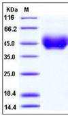
CD274 / B7-H1 / PD-L1 Protein - Recombinant  Programmed Cell Death Protein 1 Ligand 1 By SDS-PAGE
Select
E. coli
His-GST
53.7 kDa
10 µg/$286; 50 µg/$472; 100 µg/$752; 200 µg/$942; 1 mg/$2,069; 500 µg/$1,600; 5 mg/$3,291; 2 mg/$2,257
Select
Human Cells
6His, C-terminus
Less than 1.0 EU/µg protein (determined by LAL method).
25.6kD
10 µg/$248; 50 µg/$288; 500 µg/$785; 1 mg/$1,203
Select
E. coli
271 AA
His
Less than 0.1 EU/µg protein (determined by LAL method).
29.92 kDa
10 mg/$4,268; 5 mg/$3,201; 1 mg/$1,868; 0.5 mg/$1,475; 200 µg/$1,103; 100 µg/$756; 50 µg/$531
CD274 / B7-H1 / PD-L1 Protein
Select
E. coli
His
20 µg/$261; 100 µg/$323; 10 µg/$242; 250 µg/$365; 500 µg/$515
CD274 / B7-H1 / PD-L1 Protein
Select
E. coli
His
100 µg/$361; 500 µg/$739; 20 µg/$261
Select
Human Cells
6His, C-terminus
Less than 1.0 EU/µg protein (determined by LAL method).
26.33kD
10 µg/$240; 50 µg/$299; 500 µg/$929; 1 mg/$1,245
Select
Human Cells
DDK,C-terminus
Less than 1.0 EU/µg protein (determined by LAL method).
26.3kD
10 µg/$258; 50 µg/$321; 500 µg/$1,262; 1 mg/$1,627
Select
Human Cells
mFc, C-terminus
Less than 1.0 EU/µg protein (determined by LAL method).
50.9kD
10 µg/$258; 50 µg/$321; 500 µg/$1,262; 1 mg/$1,627
Select
Human Cells
Fc, C-terminus
Less than 1.0 EU/µg protein (determined by LAL method).
51.7kD
10 µg/$248; 50 µg/$288; 500 µg/$785; 1 mg/$1,203
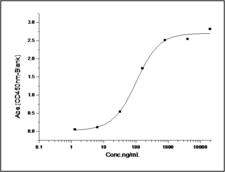
CD274 / B7-H1 / PD-L1 Protein - Measured by its binding ability in a functional ELISA. Immobilized human B7-H1 at 20 µg/ml (100 µl/well) can bind human PD1 with a linear ranger of 0.032-0.8 µg/ml.
Select
HEK 293 Cells
His,C-terminus
Bind ELISA Func SDS-PAGE
Biologically active
Less than 1.0 EU/µg protein (determined by LAL method).
The recombinant mature human B7-H1 comprises 232 amino acids and predicts a molecular mass of 26.8 kDa. As a result of glycosylation, the rh B7-H1 migrates as an approximately 35-38 kDa protein in SDS-PAGE under reducing conditions.
100 µg/$441; 1 mg/$2,003
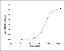
CD274 / B7-H1 / PD-L1 Protein - Measured by its binding ability in a functional ELISA. Immobilized human PD-1 at 10 µg/ml (100 µl/well) can bind recombinant human B7-H1 / PD-L1 / Fc chimera with a linear range of 0.02-0.4 µg/ml.
Select
HEK 293 Cells
None
Bind ELISA Func SDS-PAGE
Biologically active
Less than 1.0 EU/µg protein (determined by LAL method).
The recombinant mature human B7-H1/Fc is a disulfide-linked homodimeric protein. The reduced monomer consists of 459 amino acids and predicts a molecular mass of 52 kDa. As a result of glycosylation, the rh B7-H1/Fc monomer migrates as an approximately 65-70 kDa protein in SDS-PAGE under reducing conditions.
100 µg/$441; 1 mg/$2,261
Select
HEK 293 Cells
Mouse IgG1 Fc, C-terminus
Bind ELISA Func SDS-PAGE
Biologically active
Less than 1.0 EU/µg protein (determined by LAL method).
The recombinant human CD274 consists of 455 amino acids and predicts a molecular mass of 51.7 kDa.
100 µg/$372; 1 mg/$1,498
CD274 / B7-H1 / PD-L1 Protein
Select
HEK 293 Cells
His,C-terminus
Bind ELISA Func SDS-PAGE
Biologically active
Less than 1.0 EU/µg protein (determined by LAL method).
The secreted recombinant mouse CD274 comprises 231 amino acids and has a predicted molecular mass of 26.3 kDa. As a result of glycosylation, it migrates as an approximately 40-45 kDa band in SDS-PAGE under reducing conditions.
100 µg/$403; 1 mg/$1,697
Select
HEK 293 Cells
Human IgG Fc, C-terminus
Bind ELISA Func SDS-PAGE
Biologically active
Less than 1.0 EU/µg protein (determined by LAL method).
The recombinant mouse Cd274 consists 459 amino acids and predicts a molecular mass of 51.6 kDa.
100 µg/$348; 1 mg/$1,345
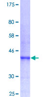
CD274 / B7-H1 / PD-L1 Protein - 12.5% SDS-PAGE Stained with Coomassie Blue.
Select
Wheat Germ Extract
GST, N-terminus
10 µg/$479; 25 µg/$670
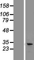
CD274 / B7-H1 / PD-L1 Protein - Western validation with an anti-DDK antibody * L: Control HEK293 lysate R: Over-expression lysate
Select
293T Cells
Myc-DDK (Flag)
33.28 kDa
20 µg/$150
Select
Human Cells
His
Less than 1.0 EU/µg protein (determined by LAL method).
26.3 kDa
100 µg/$661; 1 mg/$3,499
Select
Human Cells
Fc, C-terminus
Less than 1.0 EU/µg protein (determined by LAL method).
52.4kD
10 µg/$258; 50 µg/$321; 500 µg/$1,262; 1 mg/$1,627
CD274 / B7-H1 / PD-L1 Protein - Western validation with an anti-DDK antibody * L: Control HEK293 lysate R: Over-expression lysate
Select
HEK 293 Cells
Myc-DDK (Flag)
33.28 kDa
100 µg/$494
CD274 / B7-H1 / PD-L1 Protein
Select
HEK 293 Cells
Fc, C-terminus
50 kDa
20 µg/$1,107
CD274 / B7-H1 / PD-L1 Protein
Select
HEK 293 Cells
DDK (Flag)-His, C-terminus
20 µg/$1,107
CD274 / B7-H1 / PD-L1 Protein
Select
HEK 293 Cells
Fc, C-terminus
50 kDa
20 µg/$1,107
Select
HEK 293 Cells
6His, C-terminus
Less than 0.2 EU/µg protein (determined by LAL method).
31-35 kDa
100 µg/$338; 1 mg/$1,475
Select
HEK 293 Cells
None
Biologically active
Less than 0.01 EU/µg protein (determined by LAL method).
~35 kDa (SDS-PAGE)
50 µg/$442
Select
HEK 293 Cells
Mouse IgG2a Fc, N-terminus
Biologically active
Less than 5.0 EU/mg protein (determined by LAL method).
~70 kDa (SDS-PAGE)
100 µg/$463
Viewing 1-25 of 55 product results


Filtered By:
Products: Proteins


If you do not find the reagent or information you require, please contact Customer.Support@LSBio.com to inquire about additional products in development.

PLEASE NOTE

For RESEARCH USE ONLY. Intended for use by laboratory professionals. Not intended for human diagnostic or therapeutic purposes.

The data on this page has been compiled from LifeSpan internal sources, the National Center for Biotechnology Information (NCBI), and The Universal Protein Resource (UniProt).