Products
Research Areas
COVID-19
Resources
Login
Quick Order
Cart Cart lightblue
Login
Registration enables users to use special features of this website, such as past
order histories, retained contact details for faster checkout, review submissions, and special promotions.


Fields marked with a * are required.

Login
Quick Order
Contact Us

Location


Corporate Headquarters

Vector Laboratories, Inc.
6737 Mowry Ave
Newark, CA 94560
United States

Telephone Numbers


Customer Service: (800) 227-6666 / (650) 697-3600


Contact Us


Additional Contact Details

Login
Registration enables users to use special features of this website, such as past
order histories, retained contact details for faster checkout, review submissions, and special promotions.


Fields marked with a * are required.

Login
Quick Order

AKT2

v-akt murine thymoma viral oncogene homolog 2

AKT2 is one of 3 closely related serine/threonine-protein kinases (AKT1, AKT2 and AKT3) called the AKT kinase, and which regulate many processes including metabolism, proliferation, cell survival, growth and angiogenesis. This is mediated through serine and/or threonine phosphorylation of a range of downstream substrates. Over 100 substrate candidates have been reported so far, but for most of them, no isoform specificity has been reported. AKT is responsible of the regulation of glucose uptake by mediating insulin-induced translocation of the SLC2A4/GLUT4 glucose transporter to the cell surface. Phosphorylation of PTPN1 at 'Ser-50' negatively modulates its phosphatase activity preventing dephosphorylation of the insulin receptor and the attenuation of insulin signaling. Phosphorylation of TBC1D4 triggers the binding of this effector to inhibitory 14-3-3 proteins, which is required for insulin-stimulated glucose transport. AKT regulates also the storage of glucose in the form of glycogen by phosphorylating GSK3A at 'Ser-21' and GSK3B at 'Ser-9', resulting in inhibition of its kinase activity. Phosphorylation of GSK3 isoforms by AKT is also thought to be one mechanism by which cell proliferation is driven. AKT regulates also cell survival via the phosphorylation of MAP3K5 (apoptosis signal-related kinase). Phosphorylation of 'Ser-83' decreases MAP3K5 kinase activity stimulated by oxidative stress and thereby prevents apoptosis. AKT mediates insulin-stimulated protein synthesis by phosphorylating TSC2 at 'Ser-939' and 'Thr-1462', thereby activating mTORC1 signaling and leading to both phosphorylation of 4E-BP1 and in activation of RPS6KB1. AKT is involved in the phosphorylation of members of the FOXO factors (Forkhead family of transcription factors), leading to binding of 14-3-3 proteins and cytoplasmic localization. In particular, FOXO1 is phosphorylated at 'Thr-24', 'Ser-256' and 'Ser-319'. FOXO3 and FOXO4 are phosphorylated on equivalent sites. AKT has an important role in the regulation of NF-kappa-B-dependent gene transcription and positively regulates the activity of CREB1 (cyclic AMP (cAMP)-response element binding protein). The phosphorylation of CREB1 induces the binding of accessory proteins that are necessary for the transcription of pro-survival genes such as BCL2 and MCL1. AKT phosphorylates 'Ser-454' on ATP citrate lyase (ACLY), thereby potentially regulating ACLY activity and fatty acid synthesis. Activates the 3B isoform of cyclic nucleotide phosphodiesterase (PDE3B) via phosphorylation of 'Ser-273', resulting in reduced cyclic AMP levels and inhibition of lipolysis. Phosphorylates PIKFYVE on 'Ser-318', which results in increased PI3P-5 activity. The Rho GTPase-activating protein DLC1 is another substrate and its phosphorylation is implicated in the regulation cell proliferation and cell growth. AKT plays a role as key modulator of the AKT-mTOR signaling pathway controlling the tempo of the process of newborn neurons integration during adult neurogenesis, including correct neuron positioning, dendritic development and synapse formation. Signals downstream of phosphatidylinositol 3-kinase (PI3K) to mediate the effects of various growth factors such as platelet-derived growth factor (PDGF), epidermal growth factor (EGF), insulin and insulin-like growth factor I (IGF-I). AKT mediates the antiapoptotic effects of IGF-I. Essential for the SPATA13-mediated regulation of cell migration and adhesion assembly and disassembly. May be involved in the regulation of the placental development.One of the few specific substrates of AKT2 identified recently is PITX2. Phosphorylation of PITX2 impairs its association with the CCND1 mRNA-stabilizing complex thus shortening the half-life of CCND1. AKT2 seems also to be the principal isoform responsible of the regulation of glucose uptake. Phosphorylates C2CD5 on 'Ser-197' during insulin-stimulated adipocytes. AKT2 is also specifically involved in skeletal muscle differentiation, one of its substrates in this process being ANKRD2. Down-regulation by RNA interference reduces the expression of the phosphorylated form of BAD, resulting in the induction of caspase-dependent apoptosis. Phosphorylates CLK2 on 'Thr-343'.

Gene Name: v-akt murine thymoma viral oncogene homolog 2
Family/Subfamily: Protein Kinase , RAC/AKT
Synonyms: AKT2, PKB beta, PKBBETA, PRKBB, Protein kinase Akt-2, Protein kinase B beta, RAC-PK-beta, Rac protein kinase beta, RAC-BETA, HIHGHH, PKBB
Target Sequences: NM_001626 NP_001617.1 P31751

Publications (2)

1
Inhibition of protein kinase Akt1 by apoptosis signal-regulating kinase-1 (ASK1) is involved in apoptotic inhibition of regulatory volume increase. Subramanyam M, Takahashi N, Hasegawa Y, Mohri T, Okada Y. The Journal of biological chemistry. 2010 285:6109-17. [PubMed:20048146] [PMC:PMC2825405] Related Antibodies: LS-C19061.
2
AKT isoforms have distinct hippocampal expression and roles in synaptic plasticity. Levenga J, Wong H, Milstead RA, Keller BN, LaPlante LE, Hoeffer CA. eLife. 2017 November;6:pii: e30640. (Human) [Full Text Article] [PubMed:29173281] [PMC:PMC5722612] Related Antibodies: LS-C156232.

☰ Filters
Products
Antibodies (137)
Primary (137)
AKT2 (137)
Human (132)
Mouse (60)
Rat (41)
Bat (6)
Bovine (8)
Chicken (7)
Chimpanzee (6)
Dog (10)
Gibbon (3)
Goat (3)
Guinea pig (8)
Hamster (5)
Horse (6)
Monkey (12)
Non-Human Primate (1)
Pig (8)
Rabbit (7)
Sheep (5)
Xenopus (5)
Zebrafish (3)
IHC (72)
IHC-P (17)
WB (129)
Flo (26)
DB (10)
ELISA (79)
ICC (7)
IF (30)
IP (7)
Peptide-ELISA (5)
rabbit (93)
mouse (30)
rat (14)
Product Group
IHCPlus (2)
IgG (59)
IgG1 (19)
IgG1,k (1)
IgG2a (3)
IgG2a,k (14)
IgG2b (4)
IgG2b,k (2)
IgG3 (1)
IgM (1)
monoclonal mc (44)
polyclonal pc (93)
11F6.B2 (7)
148CT5.4.1.4 (5)
1623CT791.157.67.66 (1)
16C8.E4.G7.E9 (2)
16G11.E8 (7)
1B6 (2)
1G8C12 (1)
95C657.1.2 (1)
OTI1A2 (2)
OTI4G1 (2)
OTI4H7 (2)
OTI8D9 (2)
OTI8G8 (2)
X1 (1)
APC (1)
APC Conjugated (10)
Azide-free (8)
Biotin Conjugated (13)
Carrier-free (5)
Cy3 Conjugated (1)
Cy7 Conjugated (1)
FITC Conjugated (13)
HRP Conjugated (11)
PE Conjugated (8)
Preservative Free (1)
RPE Conjugated (2)
Unconjugated (78)
Internal (16)
aa93-123 (14)
pSer474 (13)
aa142-412 (8)
Ser474 (7)
aa214-451 (7)
aa93-122 (7)
C-Terminus (6)
aa1-481 (4)
aa71-120 (4)
400-480 aa (2)
aa2-481 (2)
aa106-123 (1)
aa272-481 (1)
aa416-444 (1)
aa431-481 (1)
aa432-481 (1)
aa450-475 (1)
aa454-481 (1)
aa455-468 (1)
p474 (1)
No (135)
Yes (2)
AKT2 Antibody - AKT2 Monoclonal Antibody immunohistochemistry of formalin-fixed and paraffin-embedded human prostate carcinoma followed by peroxidase-conjugated secondary antibody and DAB staining.
Select
AKT2 Mouse anti-Human Monoclonal (aa214-451) Antibody
Mouse, Human
IF, IHC, IHC-P, WB
Unconjugated
400 µl/$393
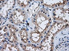
AKT2 Antibody - IHC of paraffin-embedded Kidney tissue using anti-AKT2 mouse monoclonal antibody. (Dilution 1:50).
Select
AKT2 Mouse anti-Human Monoclonal (OTI4H7) Antibody
Human
Flo, IF, IHC, IHC-P, WB
Unconjugated
100 µl/$434
AKT2 Antibody - IHC of paraffin-embedded Kidney tissue using anti-AKT2 mouse monoclonal antibody. (Dilution 1:50).
Select
AKT2 Mouse anti-Human Monoclonal (Carrier-free) (OTI4H7) Antibody
Human
Flo, IF, IHC, WB
Unconjugated, Carrier-free
100 µg/$511
Select
AKT2 Mouse anti-Human Monoclonal (aa214-451) (PE) (148CT5.4.1.4) Antibody
Mouse, Human
ELISA, ICC, IF, IHC, WB
PE Conjugated
200 µl/$952
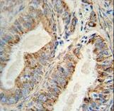
AKT2 Antibody - Formalin-fixed and paraffin-embedded human colon carcinoma reacted with AKT2 Antibody , which was peroxidase-conjugated to the secondary antibody, followed by DAB staining. This data demonstrates the use of this antibody for immunohistochemistry; clinical relevance has not been evaluated.
Select
AKT2 Rabbit anti-Human Polyclonal (aa93-123) Antibody
Mouse, Human
Flo, IF, IHC, IHC-P, WB
Unconjugated
400 µl/$393
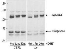
AKT2 Antibody - Anti-AKT2 Antibody - Western Blot. Anti-AKT2 Antibody in Western blot shows detection of AKT2. A lysate from a stable HEK293 cell line expressing an inducible, myristoylated form of Akt2 (MyrAkt2-ER) was loaded for SDS- PAGE, separated and then transferred to nitrocellulose. The blot was used at 1:1000 and reacted with Anti-AKT2 Antibody for 1h at room temperature. In response to 4OHT (tamoxifen), the Akt2 is recruited to the plasma membrane via its myristoylation sequence, and becomes phosphorylated and activated. The blot (right panel) shows 0 m, 15 m, and 30 m of 4OHT treatment. This treatment has no effect on endogenous Akt2, but causes a band shift upwards in the MyrAkt2. Endogenous Akt2 runs at about 60kD whereas the myristoylated construct runs at around 110kD. Antibody to AKT2 detects both unphosphorylated and phosphorylated AKT2. As before approximately 20 mg/lane of crude HEK293 lysate was loaded for SDS-PAGE, separated and then transferred to nitrocellulose. The right lane contains lysate pretreated-for-15min-with 25 uM LY294002 which affects the phosphorylation of endogenous Akt2 but has no effect on phosphorylation of the myristoylated construct. This antibody clearly detects both the phosphorylated (top arrow) and the non-phosphorylated (bottom arrow) forms of endogenous Akt2.
Select
AKT2 Rabbit anti-Human Polyclonal (aa450-475) Antibody
Human
ELISA, Flo, WB
Unconjugated
100 µg/$567
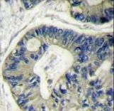
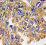
AKT2 Antibody - Formalin-fixed and paraffin-embedded human lung carcinoma tissue reacted with AKT2 Antibody (S474), which was peroxidase-conjugated to the secondary antibody, followed by DAB staining. This data demonstrates the use of this antibody for immunohistochemistry; clinical relevance has not been evaluated.
Select
AKT2 Rabbit anti-Human Polyclonal (Ser474) Antibody
Human
IF, IHC, IHC-P, WB
Unconjugated
400 µl/$393
AKT2 Antibody - Formalin-fixed and paraffin-embedded human lung carcinoma tissue reacted with AKT2 antibody , which was peroxidase-conjugated to the secondary antibody, followed by DAB staining. This data demonstrates the use of this antibody for immunohistochemistry; clinical relevance has not been evaluated.
Select
AKT2 Rabbit anti-Human Polyclonal (aa416-444) Antibody
Human
Flo, IF, IHC, IHC-P, WB
Unconjugated
400 µl/$393
Select
AKT2 Rabbit anti-Human Polyclonal (pSer474) (PE) Antibody
Human
DB, ELISA
PE Conjugated
200 µl/$952
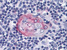
AKT2 Antibody - Anti-AKT2 antibody IHC of human thymus. Immunohistochemistry of formalin-fixed, paraffin-embedded tissue after heat-induced antigen retrieval. Antibody dilution 1:100.
Select
AKT2 Mouse anti-Human Monoclonal (1B6) Antibody
Human
ELISA, IF, IHC, IHC-P, WB
Unconjugated
50 µl/$460
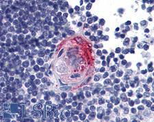
AKT2 Antibody - Human Tonsil: Formalin-Fixed, Paraffin-Embedded (FFPE)
Select
AKT2 Rabbit anti-Mouse Polyclonal (aa93-122) Antibody
Mouse, Human
IHC, IHC-P, WB
Unconjugated
200 µl/$460
AKT2 Antibody - Immunoperoxidase of monoclonal antibody to AKT2 on formalin-fixed paraffin-embedded human small Intestine. [antibody concentration 3 ug/ml]
Select
AKT2 Mouse anti-Human Monoclonal (X1) Antibody
Mouse, Rat, Human
ELISA, IF, IHC, IHC-P, WB
Unconjugated
100 µg/$423
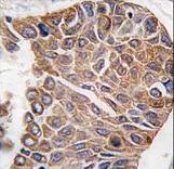
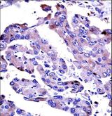
AKT2 Antibody - AKT2 Antibody immunohistochemistry of formalin-fixed and paraffin-embedded human lung adenocarcinoma followed by peroxidase-conjugated secondary antibody and DAB staining.
Select
AKT2 Rabbit anti-Human Polyclonal (aa93-123) Antibody
Human
Flo, IF, IHC, IHC-P, WB
Unconjugated
400 µl/$393
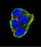
AKT2 Antibody - Confocal immunofluorescence of AKT2 Antibody with 293 cell followed by Alexa Fluor 488-conjugated goat anti-rabbit lgG (green). Actin filaments have been labeled with Alexa Fluor 555 phalloidin (red). DAPI was used to stain the cell nuclear (blue).
Select
AKT2 Rabbit anti-Human Polyclonal Antibody
Mouse, Human
Flo, IF, IHC, WB
Unconjugated
400 µl/$393
AKT2 Antibody - AKT2 Antibody in Immunohistochemistry (IHC)
Select
AKT2 Mouse anti-Human Monoclonal (1B6) Antibody
Rat, Non-Human Primate, Human
ICC, IF, IHC, WB
Unconjugated
100 µl/$518
AKT2 Antibody - Immunohistochemistry of Rat monoclonal anti AKT2 unconjugated Antibody in human breast carcinoma. Tissue: Human Breast Cancer. Fixation: FFPE buffered formalin 10% conc. Ag Retrieval: Heat, Citrate pH 6.2. Pressure Cooker. antibody: anti-AKT2 at 2ug/ml for 1.5 hour @ room temp. Secondary Ab: mouse anti-rat at 1:50 for 45” RT.
Select
AKT2 Rat anti-Human Monoclonal (Internal) (FITC) (16G11.E8) Antibody
Human
ELISA, Flo, IF, IHC, WB
FITC Conjugated
50 µg/$562
AKT2 Antibody - Immunohistochemistry of Rat monoclonal anti AKT2 unconjugated Antibody in human breast carcinoma. Tissue: Human Breast Cancer. Fixation: FFPE buffered formalin 10% conc. Ag Retrieval: Heat, Citrate pH 6.2. Pressure Cooker. antibody: anti-AKT2 at 2ug/ml for 1.5 hour @ room temp. Secondary Ab: mouse anti-rat at 1:50 for 45” RT.
Select
AKT2 Rat anti-Human Monoclonal (Internal) (Biotin) (16G11.E8) Antibody
Human
ELISA, Flo, IF, IHC, WB
Biotin Conjugated
50 µg/$562
AKT2 Antibody - Immunohistochemistry of Rat monoclonal anti AKT2 unconjugated Antibody in human breast carcinoma. Tissue: Human Breast Cancer. Fixation: FFPE buffered formalin 10% conc. Ag Retrieval: Heat, Citrate pH 6.2. Pressure Cooker. antibody: anti-AKT2 at 2ug/ml for 1.5 hour @ room temp. Secondary Ab: mouse anti-rat at 1:50 for 45” RT.
Select
AKT2 Rat anti-Human Monoclonal (Internal) (RPE) (16G11.E8) Antibody
Human
ELISA, Flo, IF, IHC, WB
RPE Conjugated
50 µg/$562
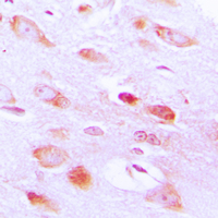
AKT2 Antibody - Immunohistochemical analysis of AKT2 staining in human brain formalin fixed paraffin embedded tissue section. The section was pre-treated using heat mediated antigen retrieval with sodium citrate buffer (pH 6.0). The section was then incubated with the antibody at room temperature and detected using an HRP conjugated compact polymer system. DAB was used as the chromogen. The section was then counterstained with hematoxylin and mounted with DPX.
Select
AKT2 Rabbit anti-Human Polyclonal (C-Terminus) Antibody
Chicken, Mouse, Dog, Rat, Human
ICC, IF, IHC, IHC-P, WB
Unconjugated
100 µl/$299
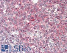
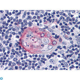
AKT2 Antibody - Immunohistochemistry (IHC) analysis of paraffin-embedded human Thymus tissues with AEC staining using Akt2 Monoclonal Antibody.
Select
AKT2 Mouse anti-Human Monoclonal Antibody
Rat, Human, Monkey
ELISA, IF, IHC, WB
Unconjugated
50 µg/$332; 100 µg/$404; 200 µg/$505
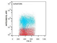
AKT2 Antibody - Flow Cytometry of Rat anti-AKT2 antibody. Cells: Jurkat Cells. Stimulation: none. Primary antibody: Allophycocyanin AKT2 antibody at 1.0 µg/mL for 20 min at 4°C.
Select
AKT2 Rat anti-Human Monoclonal (Internal) (APC) (16G11.E8) Antibody
Human
DB, ELISA, Flo, IF, IHC, WB
APC Conjugated
50 µg/$582
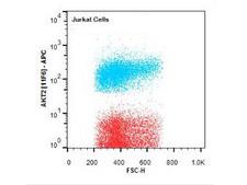
AKT2 Antibody - Flow Cytometry of Rat anti-AKT2 antibody. Cells: Jurkat Cells. Stimulation: none. Primary antibody: Allophycocyanin conjugated AKT2 antibody at 1.0 µg/mL for 20 min at 4°C.
Select
AKT2 Rat anti-Human Monoclonal (Internal) (APC) (11F6.B2) Antibody
Human
DB, ELISA, Flo, IF, IHC, WB
APC Conjugated
50 µg/$582
AKT2 Antibody - Western Blot of Rat Anti-AKT2 antibody. Lane 1: LnCap lysate Lane 2: Jurkat lysate. Lane 3: MDA-MB 468 lysate. Load: 5 µg per lane. Primary antibody: AKT2 unconjugated antibody at 1:1000 for overnight at 4°C. Secondary antibody: Rat secondary antibody at 1:20,000 for 45 min at RT. Block: 5% BLOTTO overnight at 4°C. Predicted/Observed size: 56 kDa for AKT2.
Select
AKT2 Rat anti-Human Monoclonal (Internal) (FITC) (11F6.B2) Antibody
Human
ELISA, Flo, IF, IHC, WB
FITC Conjugated
50 µg/$562
AKT2 Antibody - Western Blot of Rat Anti-AKT2 antibody. Lane 1: LnCap lysate Lane 2: Jurkat lysate. Lane 3: MDA-MB 468 lysate. Load: 5 µg per lane. Primary antibody: AKT2 unconjugated antibody at 1:1000 for overnight at 4°C. Secondary antibody: Rat secondary antibody at 1:20,000 for 45 min at RT. Block: 5% BLOTTO overnight at 4°C. Predicted/Observed size: 56 kDa for AKT2.
Select
AKT2 Rat anti-Human Monoclonal (Internal) (Biotin) (11F6.B2) Antibody
Human
ELISA, Flo, IF, IHC, WB
Biotin Conjugated
50 µg/$562
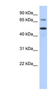
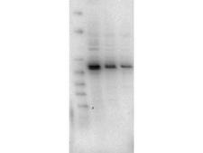
AKT2 Antibody - AKT2 antibody Western blot of MCF7 cell lysate. This image was taken for the unconjugated form of this product. Other forms have not been tested.
Select
AKT2 Rabbit anti-Human Polyclonal (C-Terminus) Antibody
Mouse, Human
WB
Unconjugated
100 µl/$424
Viewing 1-25 of 137 product results


Filtered By:
Products: Antibodies


If you do not find the reagent or information you require, please contact Customer.Support@LSBio.com to inquire about additional products in development.

PLEASE NOTE

For RESEARCH USE ONLY. Intended for use by laboratory professionals. Not intended for human diagnostic or therapeutic purposes.

The data on this page has been compiled from LifeSpan internal sources, the National Center for Biotechnology Information (NCBI), and The Universal Protein Resource (UniProt).