order histories, retained contact details for faster checkout, review submissions, and special promotions.
Forgot password?
order histories, retained contact details for faster checkout, review submissions, and special promotions.
Location
Corporate Headquarters
Vector Laboratories, Inc.
6737 Mowry Ave
Newark, CA 94560
United States
Telephone Numbers
Customer Service: (800) 227-6666 / (650) 697-3600
Contact Us
Additional Contact Details
order histories, retained contact details for faster checkout, review submissions, and special promotions.
Forgot password?
order histories, retained contact details for faster checkout, review submissions, and special promotions.
| Catalog Number | Size | Price |
|---|---|---|
| LS-J1036-10 | 10 ml | $217 |
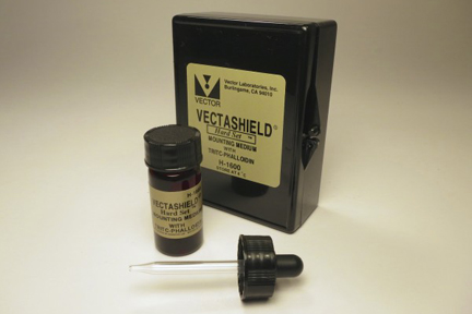
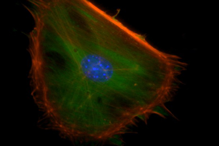
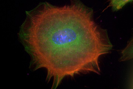
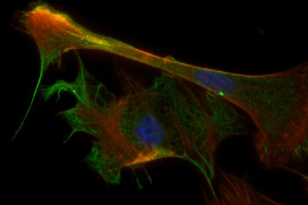


VECTASHIELD HardSet Antifade Mounting Medium with Phalloidin - LS-J1036
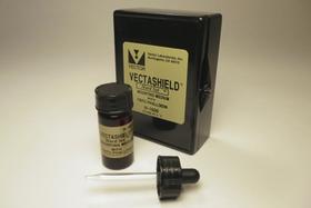
VECTASHIELD HardSet Antifade Mounting Medium with Phalloidin - LS-J1036
Overview
Specifications
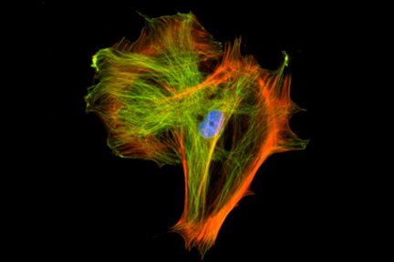
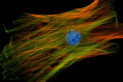
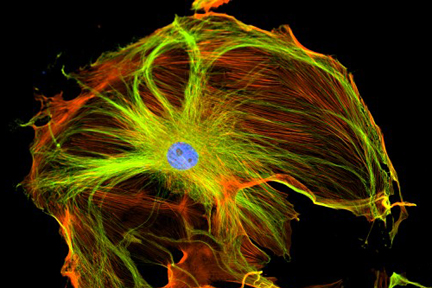
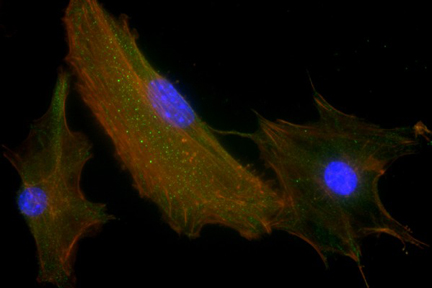
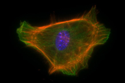
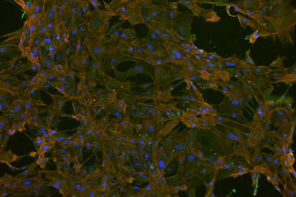
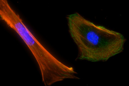
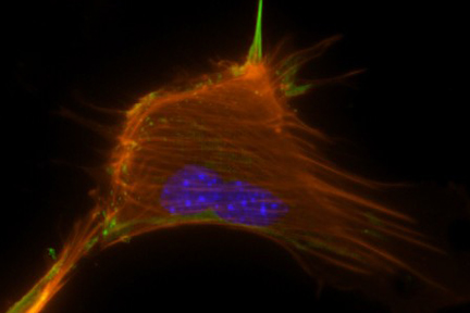
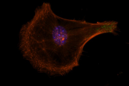
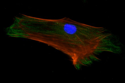
Features: - Ready-to-use aqueous solution - Contains Phalloidin counterstain (red fluorescence staining of cytoskeleton) - Hardening formulation - Inhibits photobleaching of fluorescent dyes and fluorescent proteins - No warming necessary - No sealing necessary for long term storage - Ideal refractive index (1.46) - 10 ml volume - Six month expiration date. Applications: - Immunofluorescence - In situ hybridization - Cellular Imaging - Super-Resolution Microscopy Details: VECTASHIELD HardSet Antifade Mounting Medium with Phalloidin preserves fluorescence and hardens after coverslipping. After approximately 15 minutes at room temperature, the coverslip will become immobilized, and optimal antifade ability and refractive index will be achieved. This solution contains TRITC-Phalloidin. Phalloidin is a bicyclic heptapeptide that specifically binds at the interface between the F-actin subunits. Fluorescent derivatives of Phalloidin are used to stain actin filaments. TRITC (tetramethylrhodamine) is excited at 544 nm and emits at 572 nm, producing an orange-red fluorescence. The Phalloidin concentration can be modified by mixing with VECTASHIELD HardSet Antifade Mounting Medium without Phalloidin (LS-F1034). VECTASHIELD® Mounting Media are compatible with a wide array of fluorochromes, enzymatic substrates, and fluorescent proteins. See the VECTASHIELD® Mounting Media Compatibility document to determine if VECTASHIELD® will be compatible in your system. Antifade Comparison: Other manufacturers measure the antifade properties of their mounts using labeled microspheres or arrayed spots. Vector® Labs prefers to measure antifade properties of VECTASHIELD® mounts using frozen tissue sections immunohistochemically stained with fluorescently labeled secondary antibodies. Antifade capability is measured using a 40x objective with real time imaging over 30 seconds of continuous exposure to the excitation illumination. Individual intensity measurements are recorded from 6 separate labeled regions and the average is calculated. The intensity after 30 second exposure is expressed as a percentage of the intensity at zero time. The values for PG are taken from the manufacturer s published results.

Reviews (0)
Images
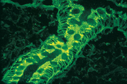
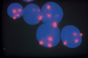
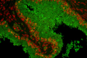
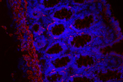
Immunofluorescence
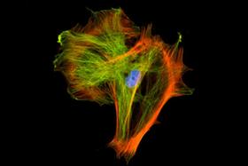
Immunofluorescence
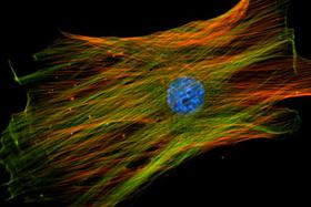
Immunofluorescence
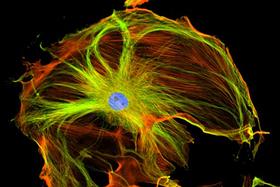
Immunofluorescence
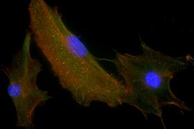

Immunofluorescence

Immunofluorescence

Immunofluorescence

Immunofluorescence


Immunofluorescence

Immunofluorescence

Immunofluorescence

Immunofluorescence


Immunofluorescence

Immunofluorescence

Immunofluorescence

Immunofluorescence

Related Products
Request SDS/MSDS
To request an SDS/MSDS form for this product, please contact our Technical Support department at:
Technical.Support@LSBio.com
