Products
Research Areas
COVID-19
Resources
Login
Quick Order
Cart Cart lightblue
Login
Registration enables users to use special features of this website, such as past
order histories, retained contact details for faster checkout, review submissions, and special promotions.


Fields marked with a * are required.

Login
Quick Order
Contact Us

Location


Corporate Headquarters

Vector Laboratories, Inc.
6737 Mowry Ave
Newark, CA 94560
United States

Telephone Numbers


Customer Service: (800) 227-6666 / (650) 697-3600


Contact Us


Additional Contact Details

Login
Registration enables users to use special features of this website, such as past
order histories, retained contact details for faster checkout, review submissions, and special promotions.


Fields marked with a * are required.

Login
Quick Order

H1F0

H1 histone family, member 0

Histones are basic nuclear proteins that are responsible for the nucleosome structure of the chromosomal fiber in eukaryotes. Nucleosomes consist of approximately 146 bp of DNA wrapped around a histone octamer composed of pairs of each of the four core histones (H2A, H2B, H3, and H4). The chromatin fiber is further compacted through the interaction of a linker histone, H1, with the DNA between the nucleosomes to form higher order chromatin structures. This gene is intronless and encodes a member of the histone H1 family.

Gene Name: H1 histone family, member 0
Synonyms: H1F0, H1 histone family, member 0, Histone H1(0), H1.0, H1(0), H1-0, H10, H1FV, Histone H1, Histone H1.0
Target Sequences: NM_005318 NP_005309.1 P07305

☰ Filters
Products
Antibodies (32)
Primary (32)
H1F0 (32)
Human (32)
Mouse (7)
Rat (6)
Bat (3)
Bovine (3)
Chimpanzee (3)
Dog (3)
Gibbon (3)
Guinea pig (3)
Hamster (3)
Monkey (4)
Orangutan (3)
Pig (3)
Xenopus (3)
IHC (12)
IHC-P (5)
WB (27)
ELISA (17)
IF (13)
IP (2)
Peptide-ELISA (3)
rabbit (28)
mouse (4)
IgG (25)
IgG2a (4)
monoclonal mc (5)
polyclonal pc (27)
Biotin Conjugated (2)
FITC Conjugated (2)
HRP Conjugated (2)
PE Conjugated (1)
Unconjugated (25)
aa36-85 (4)
aa1-194 (2)
40-120 aa, Internal (1)
Internal (1)
aa71-120 (1)
aa75-125 (1)
No (32)
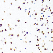
H1F0 Antibody - Immunohistochemistry of paraffin-embedded mouse brain tissue.
Select
H1F0 Rabbit anti-Human Polyclonal Antibody
Mouse, Rat, Human
IHC, WB
Unconjugated
20 µl/$287; 100 µl/$431
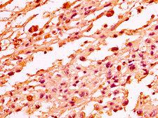
H1F0 Antibody - Immunohistochemistry image at a dilution of 1:50 and staining in paraffin-embedded human melanoma cancer performed on a Leica BondTM system. After dewaxing and hydration, antigen retrieval was mediated by high pressure in a citrate buffer (pH 6.0) . Section was blocked with 10% normal goat serum 30min at RT. Then primary antibody (1% BSA) was incubated at 4 °C overnight. The primary is detected by a biotinylated secondary antibody and visualized using an HRP conjugated SP system.
Select
H1F0 Rabbit anti-Human Polyclonal Antibody
Human
ELISA, IF, IHC, IHC-P, WB
Unconjugated
50 µl/$298; 100 µl/$370
Select
H1F0 Rabbit anti-Human Monoclonal Antibody
Human
ELISA
Unconjugated
50 µl/$316; 100 µl/$386
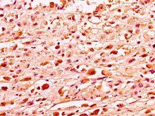
H1F0 Antibody - Immunohistochemistry image at a dilution of 1:50 and staining in paraffin-embedded human melanoma cancer performed on a Leica BondTM system. After dewaxing and hydration, antigen retrieval was mediated by high pressure in a citrate buffer (pH 6.0) . Section was blocked with 10% normal goat serum 30min at RT. Then primary antibody (1% BSA) was incubated at 4 °C overnight. The primary is detected by a biotinylated secondary antibody and visualized using an HRP conjugated SP system.
Select
H1F0 Rabbit anti-Human Polyclonal Antibody
Human
ELISA, IF, IHC, IHC-P
Unconjugated
50 µl/$298; 100 µl/$370
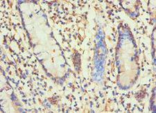
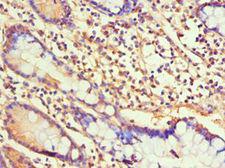
H1F0 Antibody - Immunohistochemistry of paraffin-embedded human small intestine using antibody at 1:100 dilution.
Select
H1F0 Rabbit anti-Human Polyclonal (aa1-194) Antibody
Human
ELISA, IHC, IHC-P, WB
Unconjugated
50 µl/$294; 100 µl/$360
Select
H1F0 Mouse anti-Human Monoclonal (HRP) Antibody
Human
ELISA, IF, IHC, WB
HRP Conjugated
100 µl/$973
Select
H1F0 Mouse anti-Human Monoclonal (Biotin) Antibody
Human
ELISA, IF, IHC, WB
Biotin Conjugated
100 µl/$973
Select
H1F0 Mouse anti-Human Monoclonal (FITC) Antibody
Human
ELISA, IF, IHC, WB
FITC Conjugated
100 µl/$973
Select
H1F0 Mouse anti-Human Monoclonal (PE) Antibody
Human
ELISA, IF, IHC, WB
PE Conjugated
100 µl/$973
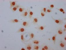
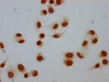
H1F0 Antibody - Immunocytochemistry analysis of Mono-methyl-H1F0 (K101) Antibody diluted at 1:5 and staining in Hela cells performed on a Leica BondTM system. The cells were fixed in 4% formaldehyde, permeabilized using 0.2% Triton X-100 and blocked with 10% normal goat serum 30min at RT. Then primary antibody (1% BSA) was incubated at 4°C overnight. The primary is detected by a biotinylated secondary antibody and visualized using an HRP conjugated SP system.
Select
H1F0 Rabbit anti-Human Polyclonal Antibody
Human
ELISA, IF, WB
Unconjugated
50 µl/$298; 100 µl/$370
Select
H1F0 Rabbit anti-Human Polyclonal Antibody
Human
ELISA, IF, IP, WB
Unconjugated
100 µg/$331
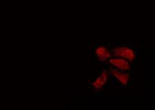
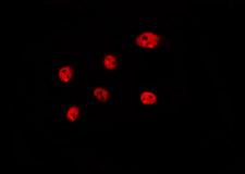
H1F0 Antibody - Staining A549 cells by IF/ICC. The samples were fixed with PFA and permeabilized in 0.1% Triton X-100, then blocked in 10% serum for 45 min at 25°C. The primary antibody was diluted at 1:200 and incubated with the sample for 1 hour at 37°C. An Alexa Fluor 594 conjugated goat anti-rabbit IgG (H+L) antibody, diluted at 1/600, was used as secondary antibody.
Select
H1F0 Rabbit anti-Human Polyclonal Antibody
Mouse, Rat, Human
IF, Peptide-ELISA, WB
Unconjugated
100 µl/$379; 200 µl/$421
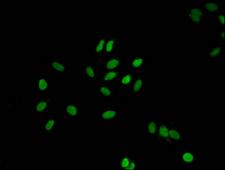
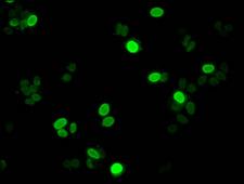
H1F0 Antibody - Immunofluorescent analysis of Hela cells at a dilution of 1:100 and Alexa Fluor 488-congugated AffiniPure Goat Anti-Rabbit IgG(H+L)
Select
H1F0 Rabbit anti-Human Polyclonal Antibody
Human
ELISA, IF, WB
Unconjugated
50 µl/$298; 100 µl/$370
H1F0 Antibody - Western blot of Histone 1.0 antibody
Select
H1F0 Rabbit anti-Human Polyclonal (40-120 aa, Internal) Antibody
Human
ELISA, IHC, WB
Unconjugated
50 µg/$295; 100 µg/$335; 200 µg/$394
H1F0 Antibody - Immunocytochemistry analysis of Mono-methyl-H1F0 (K81) Antibody diluted at 1:5 and staining in Hela cells performed on a Leica BondTM system. The cells were fixed in 4% formaldehyde, permeabilized using 0.2% Triton X-100 and blocked with 10% normal goat serum 30min at RT. Then primary antibody (1% BSA) was incubated at 4°C overnight. The primary is detected by a biotinylated secondary antibody and visualized using an HRP conjugated SP system.
Select
H1F0 Rabbit anti-Human Polyclonal Antibody
Human
ELISA, IF
Unconjugated
50 µl/$298; 100 µl/$370
H1F0 Antibody - Immunohistochemistry of paraffin-embedded human small intestine using antibody at 1:100 dilution.
Select
H1F0 Rabbit anti-Human Polyclonal (aa1-194) Antibody
Human
ELISA, IHC, IHC-P
Unconjugated
50 µl/$294; 100 µl/$360
H1F0 Antibody - Staining HeLa cells by IF/ICC. The samples were fixed with PFA and permeabilized in 0.1% Triton X-100, then blocked in 10% serum for 45 min at 25°C. The primary antibody was diluted at 1:200 and incubated with the sample for 1 hour at 37°C. An Alexa Fluor 594 conjugated goat anti-rabbit IgG (H+L) Ab, diluted at 1/600, was used as the secondary antibody.
Select
H1F0 Rabbit anti-Human Polyclonal Antibody
Human
IF, Peptide-ELISA, WB
Unconjugated
100 µl/$379; 200 µl/$421
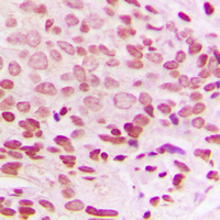
H1F0 Antibody - Immunohistochemical analysis of Histone H1.0 staining in human breast cancer formalin fixed paraffin embedded tissue section. The section was pre-treated using heat mediated antigen retrieval with sodium citrate buffer (pH 6.0). The section was then incubated with the antibody at room temperature and detected using an HRP conjugated compact polymer system. DAB was used as the chromogen. The section was then counterstained with hematoxylin and mounted with DPX.
Select
H1F0 Rabbit anti-Human Polyclonal (Internal) Antibody
Human, Monkey
IHC, IHC-P, WB
Unconjugated
100 µl/$299
H1F0 Antibody - Immunofluorescent analysis of HepG2 cells at a dilution of 1:100 and Alexa Fluor 488-congugated AffiniPure Goat Anti-Rabbit IgG(H+L)
Select
H1F0 Rabbit anti-Human Polyclonal Antibody
Human
ELISA, IF
Unconjugated
50 µl/$298; 100 µl/$370
Select
H1F0 Rabbit anti-Human Polyclonal Antibody
Mouse, Rat, Human
IHC, WB
Unconjugated
60 µl/$343; 120 µl/$427; 200 µl/$559
H1F0 Antibody - Positive Western Blot detected in Rat spleen tissue. All lanes: H1F0 antibody at 1.5 µg/ml Secondary Goat polyclonal to rabbit IgG at 1/50000 dilution. Predicted band size: 32 KDa. Observed band size: 32 KDa
Select
H1F0 Rabbit anti-Human Polyclonal Antibody
Human
ELISA, WB
Unconjugated
50 µl/$298; 100 µl/$370
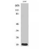
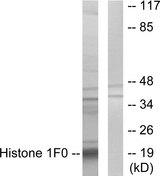
H1F0 Antibody - Western blot analysis of extracts from A549 cells, using Histone 1F0 antibody.
Select
H1F0 Rabbit anti-Human Polyclonal Antibody
Human
ELISA, WB
Unconjugated
100 µg/$357
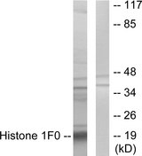
H1F0 Antibody - Western blot analysis of lysates from A549 cells, using Histone 1F0 Antibody. The lane on the right is blocked with the synthesized peptide.
Select
H1F0 Rabbit anti-Human Polyclonal (aa71-120) Antibody
Human
Peptide-ELISA, WB
Unconjugated
50 µl/$334; 100 µl/$397
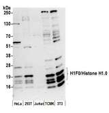
H1F0 Antibody - Detection of human and mouse H1F0/Histone H1 by western blot. Samples: Whole cell lysate (50 µg) from HeLa, HEK293T, Jurkat, mouse TCMK-1, and mouse NIH 3T3 cells prepared using NETN lysis buffer. Antibody: Affinity purified rabbit anti-H1F0/Histone H1.0 antibody used for WB at 0.4 µg/ml. Detection: Chemiluminescence with an exposure time of 3 minutes.
Select
H1F0 Rabbit anti-Human Polyclonal (aa75-125) Antibody
Mouse, Human
IP, WB
Unconjugated
10 µl/$295; 100 µl/$514
Select
H1F0 Rabbit anti-Human Polyclonal Antibody
Human
WB
Unconjugated
20 µl/$275; 100 µl/$389
Viewing 1-25 of 32 product results


Filtered By:
Products: Antibodies


If you do not find the reagent or information you require, please contact Customer.Support@LSBio.com to inquire about additional products in development.

PLEASE NOTE

For RESEARCH USE ONLY. Intended for use by laboratory professionals. Not intended for human diagnostic or therapeutic purposes.

The data on this page has been compiled from LifeSpan internal sources, the National Center for Biotechnology Information (NCBI), and The Universal Protein Resource (UniProt).