Products
Research Areas
COVID-19
Resources
Login
Quick Order
Cart Cart lightblue
Login
Registration enables users to use special features of this website, such as past
order histories, retained contact details for faster checkout, review submissions, and special promotions.


Fields marked with a * are required.

Login
Quick Order
Contact Us

Location


Corporate Headquarters

Vector Laboratories, Inc.
6737 Mowry Ave
Newark, CA 94560
United States

Telephone Numbers


Customer Service: (800) 227-6666 / (650) 697-3600


Contact Us


Additional Contact Details

Login
Registration enables users to use special features of this website, such as past
order histories, retained contact details for faster checkout, review submissions, and special promotions.


Fields marked with a * are required.

Login
Quick Order

ATXN1 / SCA1

ataxin 1

The autosomal dominant cerebellar ataxias (ADCA) are a heterogeneous group of neurodegenerative disorders characterized by progressive degeneration of the cerebellum, brain stem and spinal cord. Clinically, ADCA has been divided into three groups: ADCA types I-III. ADCAI is genetically heterogeneous, with five genetic loci, designated spinocerebellar ataxia (SCA) 1, 2, 3, 4 and 6, being assigned to five different chromosomes. ADCAII, which always presents with retinal degeneration (SCA7), and ADCAIII often referred to as the `pure' cerebellar syndrome (SCA5), are most likely homogeneous disorders. Several SCA genes have been cloned and shown to contain CAG repeats in their coding regions. ADCA is caused by the expansion of the CAG repeats, producing an elongated polyglutamine tract in the corresponding protein. The expanded repeats are variable in size and unstable, usually increasing in size when transmitted to successive generations. The function of the ataxins is not known. This locus has been mapped to chromosome 6, and it has been determined that the diseased allele contains 41-81 CAG repeats, compared to 6-39 in the normal allele, and is associated with spinocerebellar ataxia type 1 (SCA1). At least two transcript variants encoding the same protein have been found for this gene.

Gene Name: ataxin 1
Synonyms: ATXN1, ATX1, Ataxin 1, D6S504E, Ataxin-1, SCA1
Target Sequences: NM_000332 NP_000323.2 P54253

☰ Filters
Products
Antibodies (79)
Primary (79)
ATXN1 / SCA1 (79)
Human (65)
Mouse (62)
Rat (43)
Bat (1)
Bovine (1)
Drosophila (1)
Hamster (1)
Horse (1)
Monkey (2)
Pig (1)
IHC (35)
IHC-P (6)
WB (73)
Flo (6)
ELISA (19)
ICC (15)
IF (20)
IP (11)
Peptide-ELISA (5)
rabbit (45)
mouse (33)
goat (1)
Product Group
IHCPlus (2)
IgG (22)
IgG1 (16)
IgG2b (10)
IgG2b,k (7)
monoclonal mc (33)
polyclonal pc (46)
13B55 (1)
2B8A2 (1)
2F5 (2)
4C5 (1)
4C7B11 (1)
S65-37 (10)
S76-8 (10)
APC (2)
APC Conjugated (6)
Atto 390 Conjugated (2)
Atto 488 Conjugated (2)
Atto 594 Conjugated (2)
Azide-free (1)
Biotin Conjugated (6)
Cy3 Conjugated (2)
Cy7 Conjugated (2)
FITC Conjugated (6)
HRP Conjugated (4)
PE Conjugated (4)
PerCP Conjugated (2)
RPE Conjugated (2)
Unconjugated (39)
aa164-197 (9)
aa746-761 (9)
aa331-587 (8)
aa569-807 (8)
Ser776 (7)
720-800 aa (2)
C-Terminus (2)
pSer775 (2)
pSer776 (2)
aa350-400 (1)
aa586-815 (1)
aa686-699 (1)
aa742-791 (1)
aa766-816 (1)
aa778-808 (1)
No (79)
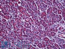
ATXN1 / SCA1 Antibody - Anti-ATXN1 / Ataxin-1 antibody IHC of human pancreas. Immunohistochemistry of formalin-fixed, paraffin-embedded tissue after heat-induced antigen retrieval. Antibody dilution 1:200.
Select
ATXN1 / SCA1 Rabbit anti-Human Polyclonal (aa742-791) Antibody
Mouse, Human
IF, IHC, IHC-P, Peptide-ELISA
Unconjugated
50 µl/$460
ATXN1 / SCA1 Antibody - Western blot of recombinant ATXN1 / Ataxin-1 / SCA1.
Select
ATXN1 / SCA1 Rabbit anti-Mouse Polyclonal (aa331-587) Antibody
Mouse
WB
Unconjugated
100 µl/$301; 200 µl/$340; 500 µl/$470; 1 ml/$535; 2 ml/$698; 5 ml/$1,161
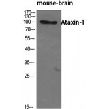
ATXN1 / SCA1 Antibody - Western blot of Ataxin-1 antibody
Select
ATXN1 / SCA1 Rabbit anti-Human Polyclonal (720-800 aa) Antibody
Mouse, Human
ELISA, IF, IHC, WB
Unconjugated
50 µg/$295; 100 µg/$335; 200 µg/$394
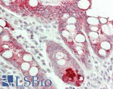
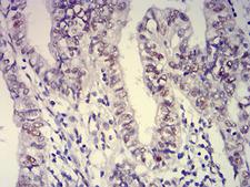
ATXN1 / SCA1 Antibody - Human Small Intestine: Formalin-Fixed, Paraffin-Embedded (FFPE)
Select
ATXN1 / SCA1 Rabbit anti-Human Polyclonal (C-Terminus) Antibody
Mouse, Rat, Human
ICC, IF, IHC, IHC-P, WB
Unconjugated
50 µl/$460
ATXN1 / SCA1 Antibody - Immunohistochemistry (IHC) analysis of paraffin-embedded ovarian cancer tissues (left) and Lung Cancer Tissues (right) with DAB staining using Ataxin-1 Monoclonal Antibody.
Select
ATXN1 / SCA1 Mouse anti-Human Monoclonal Antibody
Human
ELISA, Flo, IF, IHC, WB
Unconjugated
50 µg/$332; 100 µg/$404; 200 µg/$505
ATXN1 / SCA1 Antibody - Immunohistochemical analysis of paraffin-embedded endometrial cancer tissues using ATXN1 mouse mAb with DAB staining.
Select
ATXN1 / SCA1 Mouse anti-Human Monoclonal (4C7B11) Antibody
Mouse, Rat, Human, Monkey
ELISA, Flo, IHC, WB
Unconjugated
100 µg/$459
ATXN1 / SCA1 Antibody - Western blot using ATXN1 monoclonal antibody against HEK293 (1) and ATXN1(AA: 645-815)-hIgGFc transfected HEK293 (2) cell lysate.
Select
ATXN1 / SCA1 Mouse anti-Human Monoclonal (2F5) Antibody
Human
ELISA, Flo, IF, IHC, WB
Unconjugated
100 µl/$435
Select
ATXN1 / SCA1 Rabbit anti-Human Polyclonal Antibody
Drosophila, Rat, Mouse, Human
WB
Unconjugated
100 µl/$688
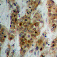
ATXN1 / SCA1 Antibody - Immunohistochemical analysis of Ataxin 1 staining in human breast cancer formalin fixed paraffin embedded tissue section. The section was pre-treated using heat mediated antigen retrieval with sodium citrate buffer (pH 6.0). The section was then incubated with the antibody at room temperature and detected using an HRP conjugated compact polymer system. DAB was used as the chromogen. The section was then counterstained with hematoxylin and mounted with DPX.
Select
ATXN1 / SCA1 Rabbit anti-Human Polyclonal (C-Terminus) Antibody
Mouse, Rat, Human
ICC, IF, IHC, IHC-P, WB
Unconjugated
30 µl/$255; 100 µl/$299; 200 µl/$351
ATXN1 / SCA1 Antibody - Western blot of Ataxin 1 expression in NIH-3T3 whole cell lysates,The lane on the left is treated with the antigen-specific peptide.
Select
ATXN1 / SCA1 Rabbit anti-Human Polyclonal Antibody
Mouse, Human
ICC, IF, IHC, WB
Unconjugated
50 µg/$305; 100 µg/$357
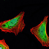
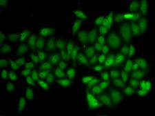
ATXN1 / SCA1 Antibody - Immunofluorescence analysis of HeLa cells.
Select
ATXN1 / SCA1 Rabbit anti-Human Polyclonal Antibody
Mouse, Rat, Human
IF, IHC, WB
Unconjugated
20 µl/$275; 100 µl/$389
Select
ATXN1 / SCA1 Mouse anti-Human Monoclonal (aa164-197) (RPE) (S76-8) Antibody
Mouse, Rat, Human
IHC, IP, WB
RPE Conjugated
100 µg/$477
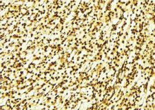
ATXN1 / SCA1 Antibody - Ataxin 1 Antibody in Immunohistochemistry (IHC)
Select
ATXN1 / SCA1 Mouse anti-Human Monoclonal (2F5) Antibody
Human
Flo, ICC, IF, IHC, WB
Unconjugated
100 µl/$541
Select
ATXN1 / SCA1 Mouse anti-Human Monoclonal (aa746-761) (PerCP) (S65-37) Antibody
Mouse, Rat, Human
ICC, WB
PerCP Conjugated
100 µg/$478
Select
ATXN1 / SCA1 Mouse anti-Human Monoclonal (13B55) Antibody
Human
ELISA, Flo, IF, IHC, WB
Unconjugated
100 µl/$688
ATXN1 / SCA1 Antibody - 1:100 staining human gastric tissue by IHC-P. The sample was formaldehyde fixed and a heat mediated antigen retrieval step in citrate buffer was performed. The sample was then blocked and incubated with the antibody for 1.5 hours at 22°C. An HRP conjugated goat anti-rabbit antibody was used as the secondary.
Select
ATXN1 / SCA1 Rabbit anti-Human Polyclonal Antibody
Mouse, Human
IF, IHC, Peptide-ELISA, WB
Unconjugated
100 µl/$379; 200 µl/$421
Select
ATXN1 / SCA1 Rabbit anti-Human Polyclonal (720-800 aa) Antibody
Mouse, Human
ELISA, IF, IHC, WB
Unconjugated
50 µg/$295; 100 µg/$335; 200 µg/$394
ATXN1 / SCA1 Antibody - Western blot analysis using *** mAb against HEK293 (1) and *** (AA: ***)-hIgGFc transfected HEK293 (2) cell lysate.
Select
ATXN1 / SCA1 Mouse anti-Human Monoclonal (2B8A2) Antibody
Mouse, Human
ELISA, Flo, WB
Unconjugated
100 µg/$459
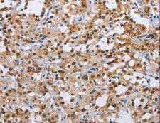
ATXN1 / SCA1 Antibody - Immunohistochemistry of paraffin-embedded Human thyroid cancer using ATXN1 Polyclonal Antibody at dilution of 1:40.
Select
ATXN1 / SCA1 Rabbit anti-Human Polyclonal Antibody
Mouse, Rat, Human
ELISA, IHC, WB
Unconjugated
20 µl/$254; 60 µl/$296; 120 µl/$355; 200 µl/$450
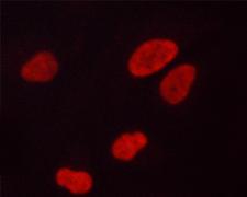
ATXN1 / SCA1 Antibody - Staining A549 cells by IF/ICC. The samples were fixed with PFA and permeabilized in 0.1% saponin prior to blocking in 10% serum for 45 min at 37°C. The primary antibody was diluted 1/400 and incubated with the sample for 1 hour at 37°C. A Alexa Fluor 594 conjugated goat polyclonal to rabbit IgG (H+L), diluted 1/600 was used as secondary antibody.
Select
ATXN1 / SCA1 Rabbit anti-Human Polyclonal (pSer775) Antibody
Mouse, Human
IF, Peptide-ELISA, WB
Unconjugated
100 µl/$379; 200 µl/$421
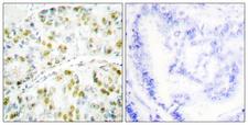
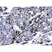
ATXN1 / SCA1 Antibody - Peptide - + Immunohistochemistry analysis of paraffin-embedded human lung carcinoma tissue using Ataxin 1 antibody.
Select
ATXN1 / SCA1 Rabbit anti-Human Polyclonal Antibody
Mouse, Human
ELISA, IF, IHC
Unconjugated
100 µg/$357
ATXN1 / SCA1 Antibody - Western blot of Ataxin 1 phosphorylation expression in Adriamycin treated HepG2 whole cell lysates,The lane on the left is treated with the antigen-specific peptide.
Select
ATXN1 / SCA1 Rabbit anti-Human Polyclonal (pSer775) Antibody
Mouse, Human
ICC, IF, WB
Unconjugated
50 µg/$305; 100 µg/$357
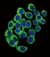
ATXN1 / SCA1 Antibody - Confocal immunofluorescence of ATXN1 Antibody (S776) with HeLa cell followed by Alexa Fluor 488-conjugated goat anti-rabbit lgG (green). DAPI was used to stain the cell nuclear (blue).
Select
ATXN1 / SCA1 Rabbit anti-Human Polyclonal (Ser776) Antibody
Human
ICC, IF, WB
Unconjugated
400 µl/$393
ATXN1 / SCA1 Antibody - Ataxin 1 antibody IHC-paraffin. IHC(P): Human Lung Cancer Tissue.
Select
ATXN1 / SCA1 Rabbit anti-Human Polyclonal (aa778-808) Antibody
Human
IHC, IHC-P, WB
Unconjugated
10 µg/$318; 100 µg/$470
Select
ATXN1 / SCA1 Mouse anti-Mouse Monoclonal Antibody
Mouse, Rat
IHC, WB
Unconjugated
100 µg/$854
Viewing 1-25 of 79 product results


Filtered By:
Products: Antibodies


If you do not find the reagent or information you require, please contact Customer.Support@LSBio.com to inquire about additional products in development.

PLEASE NOTE

For RESEARCH USE ONLY. Intended for use by laboratory professionals. Not intended for human diagnostic or therapeutic purposes.

The data on this page has been compiled from LifeSpan internal sources, the National Center for Biotechnology Information (NCBI), and The Universal Protein Resource (UniProt).