Products
Research Areas
COVID-19
Resources
Login
Quick Order
Cart Cart lightblue
Login
Registration enables users to use special features of this website, such as past
order histories, retained contact details for faster checkout, review submissions, and special promotions.


Fields marked with a * are required.

Login
Quick Order
Contact Us

Location


Corporate Headquarters

Vector Laboratories, Inc.
6737 Mowry Ave
Newark, CA 94560
United States

Telephone Numbers


Customer Service: (800) 227-6666 / (650) 697-3600


Contact Us


Additional Contact Details

Login
Registration enables users to use special features of this website, such as past
order histories, retained contact details for faster checkout, review submissions, and special promotions.


Fields marked with a * are required.

Login
Quick Order

ATP6V1A1 / ATP6V1A

ATPase, H+ transporting, lysosomal 70kDa, V1 subunit A

ATP6V1A1 / ATP6V1A is a component of vacuolar ATPase (V-ATPase), a multisubunit enzyme that mediates acidification of eukaryotic intracellular organelles. V-ATPase dependent organelle acidification is necessary for such intracellular processes as protein sorting, zymogen activation, receptor-mediated endocytosis, and synaptic vesicle proton gradient generation. V-ATPase is composed of a cytosolic V1 domain and a transmembrane V0 domain. The V1 domain consists of three A and three B subunits, two G subunits plus the C, D, E, F, and H subunits. The V1 domain contains the ATP catalytic site. The V0 domain consists of five different subunits: a, c, c', c", and d. Additional isoforms of many of the V1 and V0 subunit proteins are encoded by multiple genes or alternatively spliced transcript variants. This encoded protein is one of two V1 domain A subunit isoforms and is found in all tissues. Transcript variants derived from alternative polyadenylation exist.

Gene Name: ATPase, H+ transporting, lysosomal 70kDa, V1 subunit A
Family/Subfamily: Transporter , ATPase - V type
Synonyms: ATP6V1A, ATP6A1, ATP6V1A1, HO68, V-ATPase 69 kDa subunit, V-ATPase 69 kDa subunit 1, VA68, V-ATPase A subunit 1, Vacuolar ATPase isoform VA68, Vma1, V-ATPase subunit A, VPP2
Target Sequences: NM_001690 NP_001681.2 P38606

☰ Filters
Antibodies (21)
ELISA Kits (5)
Proteins (4)
Custom (4)
Over-Expression Lysate (2)
Primary (21)
Recombinant (2)
Sandwich (1)
ATP6V1A1 / ATP6V1A (30)
Human (21)
Mouse (10)
Rat (4)
Bovine (3)
Chicken (1)
Chimpanzee (3)
Dog (3)
Gibbon (3)
Horse (3)
Monkey (3)
Orangutan (3)
Pig (4)
Rabbit (3)
IHC (11)
IHC-P (2)
WB (20)
ELISA (9)
IP (4)
Peptide-ELISA (1)
rabbit (20)
mouse (1)
Product Group
IHCPlus (1)
IgG (15)
IgG1,k (1)
monoclonal mc (1)
polyclonal pc (20)
4F5 (1)
96-Well Strip Plate (5)
APC Conjugated (1)
Azide-free (1)
Biotin Conjugated (2)
FITC Conjugated (2)
HRP Conjugated (2)
PE Conjugated (1)
Unconjugated (13)
aa441-468 (7)
aa71-120 (4)
aa450-500 (1)
aa567-617 (1)
No (30)
Plasma (1)
Serum (1)
GST (1)
GST, N-terminus (1)
Myc-DDK (Flag) (2)
Human (3)
293T Cells (1)
HEK 293 Cells (1)
Wheat Germ Extract (2)
Purified (1)
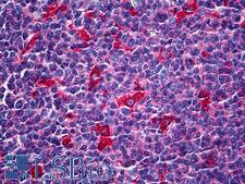
ATP6V1A1 / ATP6V1A Antibody - Anti-ATP6V1A antibody IHC of human tonsil. Immunohistochemistry of formalin-fixed, paraffin-embedded tissue after heat-induced antigen retrieval. Antibody concentration 5 ug/ml.
Select
ATP6V1A1 / ATP6V1A Mouse anti-Human Monoclonal (4F5) Antibody
Human
ELISA, IHC, IHC-P, WB
Unconjugated
50 µg/$460
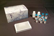
ATP6V1A1 / ATP6V1A ELISA Kit
Select
Sandwich
96-Well Strip Plate
Human
0.156 - 10 ng/ml
Colorimetric - 450nm (TMB)
Plasma, Serum
1 Plate/$669
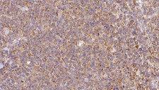
ATP6V1A1 / ATP6V1A Antibody - 1:100 staining human lymph carcinoma tissue by IHC-P. The sample was formaldehyde fixed and a heat mediated antigen retrieval step in citrate buffer was performed. The sample was then blocked and incubated with the antibody for 1.5 hours at 22°C. An HRP conjugated goat anti-rabbit antibody was used as the secondary.
Select
ATP6V1A1 / ATP6V1A Rabbit anti-Human Polyclonal Antibody
Mouse, Rat, Human
IHC, Peptide-ELISA, WB
Unconjugated
100 µl/$379; 200 µl/$421
Select
ATP6V1A1 / ATP6V1A Rabbit anti-Human Polyclonal Antibody
Human
ELISA, IHC, IP, WB
Unconjugated
100 µg/$331
Select
ATP6V1A1 / ATP6V1A Rabbit anti-Human Polyclonal Antibody
Human
ELISA, IHC, IP, WB
Unconjugated
100 µg/$341
Select
ATP6V1A1 / ATP6V1A Rabbit anti-Human Polyclonal (aa441-468) Antibody
Human
ELISA, IHC, WB
Unconjugated
200 µl/$734
Select
ATP6V1A1 / ATP6V1A Rabbit anti-Human Polyclonal (aa441-468) (Biotin) Antibody
Human
ELISA, IHC, WB
Biotin Conjugated
200 µl/$952
Select
ATP6V1A1 / ATP6V1A Rabbit anti-Human Polyclonal (aa441-468) (FITC) Antibody
Human
ELISA, IHC, WB
FITC Conjugated
200 µl/$952
Select
ATP6V1A1 / ATP6V1A Rabbit anti-Human Polyclonal (aa441-468) (APC) Antibody
Human
ELISA, IHC, WB
APC Conjugated
200 µl/$952
Select
ATP6V1A1 / ATP6V1A Rabbit anti-Human Polyclonal (aa441-468) (PE) Antibody
Human
ELISA, IHC, WB
PE Conjugated
200 µl/$952
Select
ATP6V1A1 / ATP6V1A Rabbit anti-Human Polyclonal (aa441-468) (Azide-free) (HRP) Antibody
Human
ELISA, IHC, WB
HRP Conjugated, Azide-free
200 µl/$952
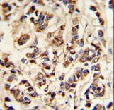
ATP6V1A1 / ATP6V1A Antibody - ATP6V1A Antibody IHC of formalin-fixed and paraffin-embedded breast carcinoma followed by peroxidase-conjugated secondary antibody and DAB staining.
Select
ATP6V1A1 / ATP6V1A Rabbit anti-Human Polyclonal (aa441-468) Antibody
Human
IHC, IHC-P, WB
Unconjugated
400 µl/$393
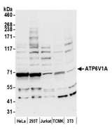
ATP6V1A1 / ATP6V1A Antibody - Detection of human and mouse ATP6V1A by western blot. Samples: Whole cell lysate (50 µg) from HeLa, HEK293T, Jurkat, mouse TCMK-1, and mouse NIH 3T3 cells prepared using NETN lysis buffer. Antibody: Affinity purified rabbit anti-ATP6V1A antibody used for WB at 0.1 µg/ml. Detection: Chemiluminescence with an exposure time of 30 seconds.
Select
ATP6V1A1 / ATP6V1A Rabbit anti-Human Polyclonal (aa567-617) Antibody
Mouse, Human
IP, WB
Unconjugated
10 µl/$295; 100 µl/$514
Select
ATP6V1A1 / ATP6V1A Rabbit anti-Mouse Polyclonal Antibody
Mouse
WB
Unconjugated
60 µl/$331; 120 µl/$403; 200 µl/$529
Select
ATP6V1A1 / ATP6V1A Rabbit anti-Human Polyclonal Antibody
Mouse, Human
WB
Unconjugated
60 µl/$331; 120 µl/$403; 200 µl/$529
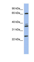
ATP6V1A1 / ATP6V1A Antibody - ATP6V1A antibody Western blot of Fetal Brain lysate. This image was taken for the unconjugated form of this product. Other forms have not been tested.
Select
ATP6V1A1 / ATP6V1A Rabbit anti-Human Polyclonal (aa71-120) Antibody
Human
WB
Unconjugated
100 µl/$424
ATP6V1A1 / ATP6V1A Antibody - ATP6V1A antibody Western blot of Fetal Brain lysate. This image was taken for the unconjugated form of this product. Other forms have not been tested.
Select
ATP6V1A1 / ATP6V1A Rabbit anti-Human Polyclonal (aa71-120) (HRP) Antibody
Rabbit, Mouse, Dog, Bovine, Pig, Horse, Rat, Chimpanzee, Orangutan, Gibbon, Human, Monkey
WB
HRP Conjugated
100 µl/$469
ATP6V1A1 / ATP6V1A Antibody - ATP6V1A antibody Western blot of Fetal Brain lysate. This image was taken for the unconjugated form of this product. Other forms have not been tested.
Select
ATP6V1A1 / ATP6V1A Rabbit anti-Human Polyclonal (aa71-120) (Biotin) Antibody
Rabbit, Mouse, Dog, Bovine, Pig, Horse, Rat, Chimpanzee, Orangutan, Gibbon, Human, Monkey
WB
Biotin Conjugated
100 µl/$469
ATP6V1A1 / ATP6V1A Antibody - ATP6V1A antibody Western blot of Fetal Brain lysate. This image was taken for the unconjugated form of this product. Other forms have not been tested.
Select
ATP6V1A1 / ATP6V1A Rabbit anti-Human Polyclonal (aa71-120) (FITC) Antibody
Rabbit, Mouse, Dog, Bovine, Pig, Horse, Rat, Chimpanzee, Orangutan, Gibbon, Human, Monkey
WB
FITC Conjugated
100 µl/$469
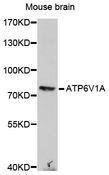
ATP6V1A1 / ATP6V1A Antibody - Western blot analysis of extracts of mouse brain , using ATP6V1A antibody at 1:1000 dilution. The secondary antibody used was an HRP Goat Anti-Rabbit IgG (H+L) at 1:10000 dilution. Lysates were loaded 25ug per lane and 3% nonfat dry milk in TBST was used for blocking. An ECL Kit was used for detection and the exposure time was 3min.
Select
ATP6V1A1 / ATP6V1A Rabbit anti-Human Polyclonal Antibody
Mouse, Human
WB
Unconjugated
20 µl/$275; 100 µl/$389
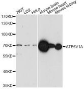
ATP6V1A1 / ATP6V1A Antibody - Western blot analysis of extracts of various cell lines, using ATP6V1A antibody at 1:1000 dilution. The secondary antibody used was an HRP Goat Anti-Rabbit IgG (H+L) at 1:10000 dilution. Lysates were loaded 25ug per lane and 3% nonfat dry milk in TBST was used for blocking. An ECL Kit was used for detection and the exposure time was 3min.
Select
ATP6V1A1 / ATP6V1A Rabbit anti-Human Polyclonal Antibody
Mouse, Human
WB
Unconjugated
20 µl/$275; 100 µl/$389
Select
ATP6V1A1 / ATP6V1A Rabbit anti-Human Polyclonal (aa450-500) Antibody
Human
IP
Unconjugated
10 µg/$295; 100 µl/$514
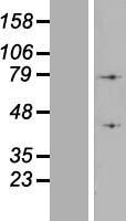
ATP6V1A1 / ATP6V1A Protein - Western validation with an anti-DDK antibody * L: Control HEK293 lysate R: Over-expression lysate
Select
293T Cells
Myc-DDK (Flag)
68.8 kDa
20 µg/$150
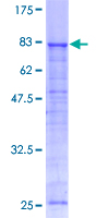
ATP6V1A1 / ATP6V1A Protein - 12.5% SDS-PAGE Stained with Coomassie Blue.
Select
Wheat Germ Extract
GST, N-terminus
10 µg/$479; 25 µg/$670
ATP6V1A1 / ATP6V1A Protein - Western validation with an anti-DDK antibody * L: Control HEK293 lysate R: Over-expression lysate
Select
HEK 293 Cells
Myc-DDK (Flag)
68.8 kDa
100 µg/$494
Viewing 1-25 of 30 product results


If you do not find the reagent or information you require, please contact Customer.Support@LSBio.com to inquire about additional products in development.

PLEASE NOTE

For RESEARCH USE ONLY. Intended for use by laboratory professionals. Not intended for human diagnostic or therapeutic purposes.

The data on this page has been compiled from LifeSpan internal sources, the National Center for Biotechnology Information (NCBI), and The Universal Protein Resource (UniProt).