Login
Registration enables users to use special features of this website, such as past
order histories, retained contact details for faster checkout, review submissions, and special promotions.
order histories, retained contact details for faster checkout, review submissions, and special promotions.
Forgot password?
Registration enables users to use special features of this website, such as past
order histories, retained contact details for faster checkout, review submissions, and special promotions.
order histories, retained contact details for faster checkout, review submissions, and special promotions.
Quick Order
Products
Antibodies
ELISA and Assay Kits
Research Areas
Infectious Disease
Resources
Purchasing
Reference Material
Contact Us
Location
Corporate Headquarters
Vector Laboratories, Inc.
6737 Mowry Ave
Newark, CA 94560
United States
Telephone Numbers
Customer Service: (800) 227-6666 / (650) 697-3600
Contact Us
Additional Contact Details
Login
Registration enables users to use special features of this website, such as past
order histories, retained contact details for faster checkout, review submissions, and special promotions.
order histories, retained contact details for faster checkout, review submissions, and special promotions.
Forgot password?
Registration enables users to use special features of this website, such as past
order histories, retained contact details for faster checkout, review submissions, and special promotions.
order histories, retained contact details for faster checkout, review submissions, and special promotions.
Quick Order
| Catalog Number | Size | Price |
|---|---|---|
| LS-G3729-50 | 50 µg | $456 |
![NAMPT / Visfatin Protein - Measurement of NAMPT enzymatic activity was performed as described previously [G.C. Elliott, et al.; Anal Biochem 107, 199 (1980)]. The recombinant Nampt was diluted in assay buffer and 10 ul per 50 ul reaction mix were applied in the reaction mix (20 mmol/l Tris-HCl pH 7.4; 2,5 mmol/l ATP; 50 mmol/l NaCl; 12,5 mmol/l MgCl2; 2 mmol/l DTT; 0,5 mmol/l PRPP; 5 uMol/l 14C-nicotinamide) and incubated at 37°C for 1h. The 50 ul reaction mix was transferred into tubes containing 2ml of acetone and afterwards pipetted onto acetone-presoaked glass microfibre filters (GF/A 24 mm). After rinsing with 2 x 1ml acetone, filters were dried, transferred into vials with 6ml scintillation cocktail and radioactivity of 14C-NMN was quantified in a liquid scintillation counter. After subtraction of buffer values as background, cpm were normalized to 10^6 cells and the volume of enzyme preparation (10 ul). Mouse liver lysate at a concentration of 34.5 ug/ml was used as positive control in each assay. The positive control is mouse liver lysate at a concentration of 34,5 ug/ml - normally, which brings the most counts per minute (cpm) (contributed by Antje Garten and Dr. Kiess, University of Leipzig, Germany).](https://lsbio-7d62.kxcdn.com/image2/human-nampt-visfatin-protein-recombinant-his-aa1-491-ls-g3729/309082_2284333.jpg)


![NAMPT / Visfatin Protein - Measurement of NAMPT enzymatic activity was performed as described previously [G.C. Elliott, et al.; Anal Biochem 107, 199 (1980)]. The recombinant Nampt was diluted in assay buffer and 10 ul per 50 ul reaction mix were applied in the reaction mix (20 mmol/l Tris-HCl pH 7.4; 2,5 mmol/l ATP; 50 mmol/l NaCl; 12,5 mmol/l MgCl2; 2 mmol/l DTT; 0,5 mmol/l PRPP; 5 uMol/l 14C-nicotinamide) and incubated at 37°C for 1h. The 50 ul reaction mix was transferred into tubes containing 2ml of acetone and afterwards pipetted onto acetone-presoaked glass microfibre filters (GF/A 24 mm). After rinsing with 2 x 1ml acetone, filters were dried, transferred into vials with 6ml scintillation cocktail and radioactivity of 14C-NMN was quantified in a liquid scintillation counter. After subtraction of buffer values as background, cpm were normalized to 10^6 cells and the volume of enzyme preparation (10 ul). Mouse liver lysate at a concentration of 34.5 ug/ml was used as positive control in each assay. The positive control is mouse liver lysate at a concentration of 34,5 ug/ml - normally, which brings the most counts per minute (cpm) (contributed by Antje Garten and Dr. Kiess, University of Leipzig, Germany).](https://lsbio-7d62.kxcdn.com/image2/human-nampt-visfatin-protein-recombinant-his-aa1-491-ls-g3729/309082_2284333.jpg)
1 of 3
2 of 3
3 of 3
Human NAMPT / Visfatin Protein (Recombinant His) (aa1-491) - LS-G3729
Human NAMPT / Visfatin Protein (Recombinant His) (aa1-491) - LS-G3729
Description:
NAMPT / Visfatin Protein LS-G3729 is a Recombinant Human NAMPT / Visfatin produced in E. coli aa 1-491 with His tag(s). It is low in endotoxin; Less than 1.0 EU/µg protein (determined by LAL method). For Research Use Only
Toll Free North America
 (800) 227-6666
For Research Use Only
Overview
Description:
NAMPT / Visfatin Protein LS-G3729 is a Recombinant Human NAMPT / Visfatin produced in E. coli aa 1-491 with His tag(s). It is low in endotoxin; Less than 1.0 EU/µg protein (determined by LAL method). For Research Use Only
Specifications
Type
Recombinant Protein
Target
NAMPT / Visfatin
Synonyms
NAMPT | PBEF | Pre-B cell-enhancing factor | VISFATIN | VF | NAmPRTase | PBEF1
Species
Human
Modifications
Unmodified
Conjugations
Unconjugated
Tag
His
Region
aa 1-491
Predicted Molecular Weight
~52kDa (SDS-PAGE)
Expression System
E. coli
Source Species
E. coli
Purification
Greater than 90% by SDS-PAGE
Bio-Activity
Shows adipogenic effects in stimulated differentiating 3T3-L1 cells.
Endotoxin
Less than 1.0 EU/µg protein (determined by LAL method).
Presentation
Lyophilized, Salt free.
Reconstitution
Reconstitute with distilled water.
Storage
Store lyophilized at -20°C for up to 1 year. Once reconstituted store at 4°C for immediate use, or aliquot and store at -20°C for up to 3 months. Avoid freeze-thaw cycles.
Restrictions
For research use only. Intended for use by laboratory professionals.
About NAMPT / Visfatin
Publications (0)
Customer Reviews (0)
Images
Functional Assay
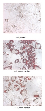
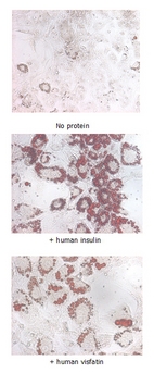
Insulin-mimetic effects on stimulated differentiating 3T3-L1 cells. 10 ug/ml iNampt (human) (rec.) (His) or human insulin was added to differentiating 3T3-L1 cells that had been stimulated with 1 uM dexamethasone and 0.5mM IBMX for 2 days. After 5 days, fat droplets were stained with Oil-Red O.
Functional Assay
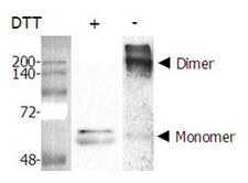
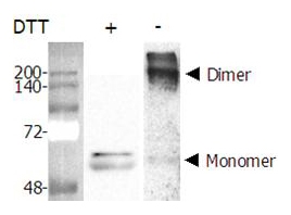
Dimer formation of recombinant Nampt (Visfatin, PBEF) (AG-40A-0018). Purified Nampt (Visfatin, PBEF) was separated by SDS-PAGE and Western blot analysis was performed using rabbit anti-Nampt polyclonal antibody (AG-25A-0025). In the absence of DTT, Nampt (visfatin, PBEF) formed a homodimer, a homomultimer.
Functional Assay
![NAMPT / Visfatin Protein - Measurement of NAMPT enzymatic activity was performed as described previously [G.C. Elliott, et al.; Anal Biochem 107, 199 (1980)]. The recombinant Nampt was diluted in assay buffer and 10 ul per 50 ul reaction mix were applied in the reaction mix (20 mmol/l Tris-HCl pH 7.4; 2,5 mmol/l ATP; 50 mmol/l NaCl; 12,5 mmol/l MgCl2; 2 mmol/l DTT; 0,5 mmol/l PRPP; 5 uMol/l 14C-nicotinamide) and incubated at 37°C for 1h. The 50 ul reaction mix was transferred into tubes containing 2ml of acetone and afterwards pipetted onto acetone-presoaked glass microfibre filters (GF/A 24 mm). After rinsing with 2 x 1ml acetone, filters were dried, transferred into vials with 6ml scintillation cocktail and radioactivity of 14C-NMN was quantified in a liquid scintillation counter. After subtraction of buffer values as background, cpm were normalized to 10^6 cells and the volume of enzyme preparation (10 ul). Mouse liver lysate at a concentration of 34.5 ug/ml was used as positive control in each assay. The positive control is mouse liver lysate at a concentration of 34,5 ug/ml - normally, which brings the most counts per minute (cpm) (contributed by Antje Garten and Dr. Kiess, University of Leipzig, Germany).](https://lsbio-7d62.kxcdn.com/image2/human-nampt-visfatin-protein-recombinant-his-aa1-491-ls-g3729/309082_5177669.jpg)
Measurement of NAMPT enzymatic activity was performed as described previously [G.C. Elliott, et al.; Anal Biochem 107, 199 (1980)]. The recombinant Nampt was diluted in assay buffer and 10 ul per 50 ul reaction mix were applied in the reaction mix (20 mmol/l Tris-HCl pH 7.4; 2,5 mmol/l ATP; 50 mmol/l NaCl; 12,5 mmol/l MgCl2; 2 mmol/l DTT; 0,5 mmol/l PRPP; 5 uMol/l 14C-nicotinamide) and incubated at 37°C for 1h. The 50 ul reaction mix was transferred into tubes containing 2ml of acetone and afterwards pipetted onto acetone-presoaked glass microfibre filters (GF/A 24 mm). After rinsing with 2 x 1ml acetone, filters were dried, transferred into vials with 6ml scintillation cocktail and radioactivity of 14C-NMN was quantified in a liquid scintillation counter. After subtraction of buffer values as background, cpm were normalized to 10^6 cells and the volume of enzyme preparation (10 ul). Mouse liver lysate at a concentration of 34.5 ug/ml was used as positive control in each assay. The positive control is mouse liver lysate at a concentration of 34,5 ug/ml - normally, which brings the most counts per minute (cpm) (contributed by Antje Garten and Dr. Kiess, University of Leipzig, Germany).
Functional Assay

Insulin-mimetic effects on stimulated differentiating 3T3-L1 cells. 10 ug/ml iNampt (human) (rec.) (His) or human insulin was added to differentiating 3T3-L1 cells that had been stimulated with 1 uM dexamethasone and 0.5mM IBMX for 2 days. After 5 days, fat droplets were stained with Oil-Red O.
Functional Assay

Dimer formation of recombinant Nampt (Visfatin, PBEF) (AG-40A-0018). Purified Nampt (Visfatin, PBEF) was separated by SDS-PAGE and Western blot analysis was performed using rabbit anti-Nampt polyclonal antibody (AG-25A-0025). In the absence of DTT, Nampt (visfatin, PBEF) formed a homodimer, a homomultimer.
Functional Assay
![NAMPT / Visfatin Protein - Measurement of NAMPT enzymatic activity was performed as described previously [G.C. Elliott, et al.; Anal Biochem 107, 199 (1980)]. The recombinant Nampt was diluted in assay buffer and 10 ul per 50 ul reaction mix were applied in the reaction mix (20 mmol/l Tris-HCl pH 7.4; 2,5 mmol/l ATP; 50 mmol/l NaCl; 12,5 mmol/l MgCl2; 2 mmol/l DTT; 0,5 mmol/l PRPP; 5 uMol/l 14C-nicotinamide) and incubated at 37°C for 1h. The 50 ul reaction mix was transferred into tubes containing 2ml of acetone and afterwards pipetted onto acetone-presoaked glass microfibre filters (GF/A 24 mm). After rinsing with 2 x 1ml acetone, filters were dried, transferred into vials with 6ml scintillation cocktail and radioactivity of 14C-NMN was quantified in a liquid scintillation counter. After subtraction of buffer values as background, cpm were normalized to 10^6 cells and the volume of enzyme preparation (10 ul). Mouse liver lysate at a concentration of 34.5 ug/ml was used as positive control in each assay. The positive control is mouse liver lysate at a concentration of 34,5 ug/ml - normally, which brings the most counts per minute (cpm) (contributed by Antje Garten and Dr. Kiess, University of Leipzig, Germany).](https://lsbio-7d62.kxcdn.com/image2/human-nampt-visfatin-protein-recombinant-his-aa1-491-ls-g3729/309082_2284333.jpg)
Measurement of NAMPT enzymatic activity was performed as described previously [G.C. Elliott, et al.; Anal Biochem 107, 199 (1980)]. The recombinant Nampt was diluted in assay buffer and 10 ul per 50 ul reaction mix were applied in the reaction mix (20 mmol/l Tris-HCl pH 7.4; 2,5 mmol/l ATP; 50 mmol/l NaCl; 12,5 mmol/l MgCl2; 2 mmol/l DTT; 0,5 mmol/l PRPP; 5 uMol/l 14C-nicotinamide) and incubated at 37°C for 1h. The 50 ul reaction mix was transferred into tubes containing 2ml of acetone and afterwards pipetted onto acetone-presoaked glass microfibre filters (GF/A 24 mm). After rinsing with 2 x 1ml acetone, filters were dried, transferred into vials with 6ml scintillation cocktail and radioactivity of 14C-NMN was quantified in a liquid scintillation counter. After subtraction of buffer values as background, cpm were normalized to 10^6 cells and the volume of enzyme preparation (10 ul). Mouse liver lysate at a concentration of 34.5 ug/ml was used as positive control in each assay. The positive control is mouse liver lysate at a concentration of 34,5 ug/ml - normally, which brings the most counts per minute (cpm) (contributed by Antje Garten and Dr. Kiess, University of Leipzig, Germany).
Functional Assay
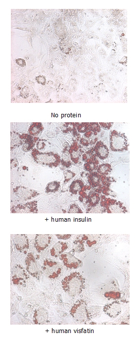
Insulin-mimetic effects on stimulated differentiating 3T3-L1 cells. 10 ug/ml iNampt (human) (rec.) (His) or human insulin was added to differentiating 3T3-L1 cells that had been stimulated with 1 uM dexamethasone and 0.5mM IBMX for 2 days. After 5 days, fat droplets were stained with Oil-Red O.
Functional Assay

Dimer formation of recombinant Nampt (Visfatin, PBEF) (AG-40A-0018). Purified Nampt (Visfatin, PBEF) was separated by SDS-PAGE and Western blot analysis was performed using rabbit anti-Nampt polyclonal antibody (AG-25A-0025). In the absence of DTT, Nampt (visfatin, PBEF) formed a homodimer, a homomultimer.
Functional Assay
![NAMPT / Visfatin Protein - Measurement of NAMPT enzymatic activity was performed as described previously [G.C. Elliott, et al.; Anal Biochem 107, 199 (1980)]. The recombinant Nampt was diluted in assay buffer and 10 ul per 50 ul reaction mix were applied in the reaction mix (20 mmol/l Tris-HCl pH 7.4; 2,5 mmol/l ATP; 50 mmol/l NaCl; 12,5 mmol/l MgCl2; 2 mmol/l DTT; 0,5 mmol/l PRPP; 5 uMol/l 14C-nicotinamide) and incubated at 37°C for 1h. The 50 ul reaction mix was transferred into tubes containing 2ml of acetone and afterwards pipetted onto acetone-presoaked glass microfibre filters (GF/A 24 mm). After rinsing with 2 x 1ml acetone, filters were dried, transferred into vials with 6ml scintillation cocktail and radioactivity of 14C-NMN was quantified in a liquid scintillation counter. After subtraction of buffer values as background, cpm were normalized to 10^6 cells and the volume of enzyme preparation (10 ul). Mouse liver lysate at a concentration of 34.5 ug/ml was used as positive control in each assay. The positive control is mouse liver lysate at a concentration of 34,5 ug/ml - normally, which brings the most counts per minute (cpm) (contributed by Antje Garten and Dr. Kiess, University of Leipzig, Germany).](https://lsbio-7d62.kxcdn.com/image2/human-nampt-visfatin-protein-recombinant-his-aa1-491-ls-g3729/309082_2284333.jpg)
Measurement of NAMPT enzymatic activity was performed as described previously [G.C. Elliott, et al.; Anal Biochem 107, 199 (1980)]. The recombinant Nampt was diluted in assay buffer and 10 ul per 50 ul reaction mix were applied in the reaction mix (20 mmol/l Tris-HCl pH 7.4; 2,5 mmol/l ATP; 50 mmol/l NaCl; 12,5 mmol/l MgCl2; 2 mmol/l DTT; 0,5 mmol/l PRPP; 5 uMol/l 14C-nicotinamide) and incubated at 37°C for 1h. The 50 ul reaction mix was transferred into tubes containing 2ml of acetone and afterwards pipetted onto acetone-presoaked glass microfibre filters (GF/A 24 mm). After rinsing with 2 x 1ml acetone, filters were dried, transferred into vials with 6ml scintillation cocktail and radioactivity of 14C-NMN was quantified in a liquid scintillation counter. After subtraction of buffer values as background, cpm were normalized to 10^6 cells and the volume of enzyme preparation (10 ul). Mouse liver lysate at a concentration of 34.5 ug/ml was used as positive control in each assay. The positive control is mouse liver lysate at a concentration of 34,5 ug/ml - normally, which brings the most counts per minute (cpm) (contributed by Antje Garten and Dr. Kiess, University of Leipzig, Germany).
Functional Assay

Insulin-mimetic effects on stimulated differentiating 3T3-L1 cells. 10 ug/ml iNampt (human) (rec.) (His) or human insulin was added to differentiating 3T3-L1 cells that had been stimulated with 1 uM dexamethasone and 0.5mM IBMX for 2 days. After 5 days, fat droplets were stained with Oil-Red O.
Functional Assay

Dimer formation of recombinant Nampt (Visfatin, PBEF) (AG-40A-0018). Purified Nampt (Visfatin, PBEF) was separated by SDS-PAGE and Western blot analysis was performed using rabbit anti-Nampt polyclonal antibody (AG-25A-0025). In the absence of DTT, Nampt (visfatin, PBEF) formed a homodimer, a homomultimer.
Functional Assay
![NAMPT / Visfatin Protein - Measurement of NAMPT enzymatic activity was performed as described previously [G.C. Elliott, et al.; Anal Biochem 107, 199 (1980)]. The recombinant Nampt was diluted in assay buffer and 10 ul per 50 ul reaction mix were applied in the reaction mix (20 mmol/l Tris-HCl pH 7.4; 2,5 mmol/l ATP; 50 mmol/l NaCl; 12,5 mmol/l MgCl2; 2 mmol/l DTT; 0,5 mmol/l PRPP; 5 uMol/l 14C-nicotinamide) and incubated at 37°C for 1h. The 50 ul reaction mix was transferred into tubes containing 2ml of acetone and afterwards pipetted onto acetone-presoaked glass microfibre filters (GF/A 24 mm). After rinsing with 2 x 1ml acetone, filters were dried, transferred into vials with 6ml scintillation cocktail and radioactivity of 14C-NMN was quantified in a liquid scintillation counter. After subtraction of buffer values as background, cpm were normalized to 10^6 cells and the volume of enzyme preparation (10 ul). Mouse liver lysate at a concentration of 34.5 ug/ml was used as positive control in each assay. The positive control is mouse liver lysate at a concentration of 34,5 ug/ml - normally, which brings the most counts per minute (cpm) (contributed by Antje Garten and Dr. Kiess, University of Leipzig, Germany).](https://lsbio-7d62.kxcdn.com/image2/human-nampt-visfatin-protein-recombinant-his-aa1-491-ls-g3729/309082_2284333.jpg)
Measurement of NAMPT enzymatic activity was performed as described previously [G.C. Elliott, et al.; Anal Biochem 107, 199 (1980)]. The recombinant Nampt was diluted in assay buffer and 10 ul per 50 ul reaction mix were applied in the reaction mix (20 mmol/l Tris-HCl pH 7.4; 2,5 mmol/l ATP; 50 mmol/l NaCl; 12,5 mmol/l MgCl2; 2 mmol/l DTT; 0,5 mmol/l PRPP; 5 uMol/l 14C-nicotinamide) and incubated at 37°C for 1h. The 50 ul reaction mix was transferred into tubes containing 2ml of acetone and afterwards pipetted onto acetone-presoaked glass microfibre filters (GF/A 24 mm). After rinsing with 2 x 1ml acetone, filters were dried, transferred into vials with 6ml scintillation cocktail and radioactivity of 14C-NMN was quantified in a liquid scintillation counter. After subtraction of buffer values as background, cpm were normalized to 10^6 cells and the volume of enzyme preparation (10 ul). Mouse liver lysate at a concentration of 34.5 ug/ml was used as positive control in each assay. The positive control is mouse liver lysate at a concentration of 34,5 ug/ml - normally, which brings the most counts per minute (cpm) (contributed by Antje Garten and Dr. Kiess, University of Leipzig, Germany).
Popular NAMPT / Visfatin Proteins
Source:
E. coli
Tag:
His-T7, N-terminus
Source:
E. coli
Tag:
His, N-Terminal
Source:
E. coli
Tag:
His, N-Terminal
Request SDS/MSDS
To request an SDS/MSDS form for this product, please contact our Technical Support department at:
Technical.Support@LSBio.com
Requested From: United States
Date Requested: 4/18/2025
Date Requested: 4/18/2025


