Login
Registration enables users to use special features of this website, such as past
order histories, retained contact details for faster checkout, review submissions, and special promotions.
order histories, retained contact details for faster checkout, review submissions, and special promotions.
Forgot password?
Registration enables users to use special features of this website, such as past
order histories, retained contact details for faster checkout, review submissions, and special promotions.
order histories, retained contact details for faster checkout, review submissions, and special promotions.
Quick Order
Products
Antibodies
ELISA and Assay Kits
Research Areas
Infectious Disease
Resources
Purchasing
Reference Material
Contact Us
Location
Corporate Headquarters
Vector Laboratories, Inc.
6737 Mowry Ave
Newark, CA 94560
United States
Telephone Numbers
Customer Service: (800) 227-6666 / (650) 697-3600
Contact Us
Additional Contact Details
Login
Registration enables users to use special features of this website, such as past
order histories, retained contact details for faster checkout, review submissions, and special promotions.
order histories, retained contact details for faster checkout, review submissions, and special promotions.
Forgot password?
Registration enables users to use special features of this website, such as past
order histories, retained contact details for faster checkout, review submissions, and special promotions.
order histories, retained contact details for faster checkout, review submissions, and special promotions.
Quick Order
| Catalog Number | Size | Price |
|---|---|---|
| LS-C392572-200 | 200 µl (0.5 mg/ml) | $393 |
1 of 3
2 of 3
3 of 3
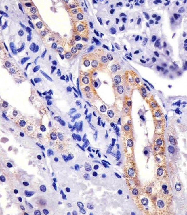
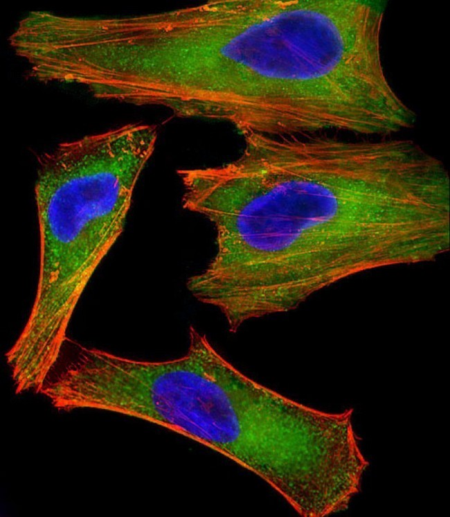
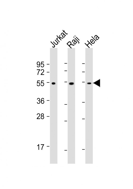
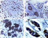
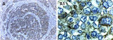
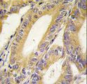
Monoclonal Mouse anti‑Human XIAP Antibody (clone 1020CT7.2.2, IHC, IF, WB) LS‑C392572
Monoclonal Mouse anti‑Human XIAP Antibody (clone 1020CT7.2.2, IHC, IF, WB) LS‑C392572
Antibody:
XIAP Mouse anti-Human Monoclonal (1020CT7.2.2) Antibody
Application:
IHC, IHC-P, IF, WB, Peptide-ELISA
Reactivity:
Human
Format:
Unconjugated, Unmodified
Toll Free North America
 (800) 227-6666
For Research Use Only
Overview
Antibody:
XIAP Mouse anti-Human Monoclonal (1020CT7.2.2) Antibody
Application:
IHC, IHC-P, IF, WB, Peptide-ELISA
Reactivity:
Human
Format:
Unconjugated, Unmodified
Specifications
Description
XIAP antibody LS-C392572 is an unconjugated mouse monoclonal antibody to human XIAP. Validated for IF, IHC, Peptide-ELISA and WB.
Target
Human XIAP
Synonyms
XIAP | API3 | BIRC4 | HILP | IAP-like protein | IAP3 | HIAP-3 | HIAP3 | IAP-3 | ILP | ILP1 | MIHA | X-linked IAP | XLP2
Host
Mouse
Reactivity
Human
(tested or 100% immunogen sequence identity)
Clonality
IgG1,k
Monoclonal
Clone
1020CT7.2.2
Conjugations
Unconjugated
Purification
Protein G purified
Modifications
Unmodified
Applications
- IHC
- IHC - Paraffin (1:25)
- Immunofluorescence (1:25)
- Western blot (1:1000 - 1:4000)
- Peptide Enzyme-Linked Immunosorbent Assay

|
Performing IHC? See our complete line of Immunohistochemistry Reagents including antigen retrieval solutions, blocking agents
ABC Detection Kits and polymers, biotinylated secondary antibodies, substrates and more.
|
Presentation
PBS, 0.09% Sodium Azide
Storage
Maintain refrigerated at 2°C to 8°C for up to 6 months. For long term storage store at -20°C.
Restrictions
For research use only. Intended for use by laboratory professionals.
About XIAP
Publications (0)
Customer Reviews (0)
Featured Products
Species:
Human
Applications:
IHC, IHC - Paraffin, ICC, Immunofluorescence, Western blot
Species:
Human, Mouse, Rat, Gerbil
Applications:
IHC, IHC - Paraffin, IHC - Frozen, Western blot, Immunoprecipitation
Species:
Human
Applications:
IHC, IHC - Paraffin, IHC - Frozen, Western blot, Immunoprecipitation
Species:
Human, Mouse
Applications:
IHC, IHC - Paraffin, Western blot
Source:
E. coli
Tag:
His, N-Terminal
Request SDS/MSDS
To request an SDS/MSDS form for this product, please contact our Technical Support department at:
Technical.Support@LSBio.com
Requested From: United States
Date Requested: 4/4/2025
Date Requested: 4/4/2025


