Login
Registration enables users to use special features of this website, such as past
order histories, retained contact details for faster checkout, review submissions, and special promotions.
order histories, retained contact details for faster checkout, review submissions, and special promotions.
Forgot password?
Registration enables users to use special features of this website, such as past
order histories, retained contact details for faster checkout, review submissions, and special promotions.
order histories, retained contact details for faster checkout, review submissions, and special promotions.
Quick Order
Products
Antibodies
ELISA and Assay Kits
Research Areas
Infectious Disease
Resources
Purchasing
Reference Material
Contact Us
Location
Corporate Headquarters
Vector Laboratories, Inc.
6737 Mowry Ave
Newark, CA 94560
United States
Telephone Numbers
Customer Service: (800) 227-6666 / (650) 697-3600
Contact Us
Additional Contact Details
Login
Registration enables users to use special features of this website, such as past
order histories, retained contact details for faster checkout, review submissions, and special promotions.
order histories, retained contact details for faster checkout, review submissions, and special promotions.
Forgot password?
Registration enables users to use special features of this website, such as past
order histories, retained contact details for faster checkout, review submissions, and special promotions.
order histories, retained contact details for faster checkout, review submissions, and special promotions.
Quick Order
| Catalog Number | Size | Price |
|---|---|---|
| LS-C751415-100 | 100 µg (1 mg/ml) | $574 |


1 of 2
2 of 2
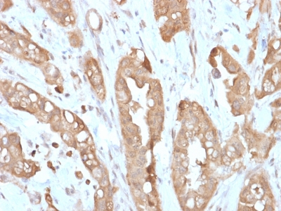
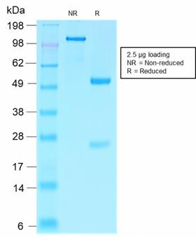
Recombinant Monoclonal Rabbit anti‑Human VIL1 / Villin Antibody (clone VIL1/2310R, Azide‑free,Carrier‑free, aa179‑311, IHC) LS‑C751415
Recombinant Monoclonal Rabbit anti‑Human VIL1 / Villin Antibody (clone VIL1/2310R, Azide‑free,Carrier‑free, aa179‑311, IHC) LS‑C751415
Antibody:
VIL1 / Villin Rabbit anti-Human Recombinant Monoclonal (aa179-311) (Azide-free, Carrier-free) (VIL1/2310R) Antibody
Application:
IHC-P
Reactivity:
Human
Format:
Unconjugated, Azide-free, Carrier-free
Other formats:
Toll Free North America
 (800) 227-6666
For Research Use Only
Overview
Antibody:
VIL1 / Villin Rabbit anti-Human Recombinant Monoclonal (aa179-311) (Azide-free, Carrier-free) (VIL1/2310R) Antibody
Application:
IHC-P
Reactivity:
Human
Format:
Unconjugated, Azide-free, Carrier-free
Other formats:
Specifications
Description
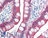
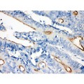
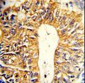
Villin antibody LS-C751415 is an unconjugated rabbit recombinant monoclonal antibody to human Villin (VIL1) (aa179-311). Validated for IHC.
Target
Human VIL1 / Villin
Synonyms
VIL1 | D2S1471 | Villin | Villin 1 | Villin-1 | VIL
Host
Rabbit
Reactivity
Human
(tested or 100% immunogen sequence identity)
Clonality
IgG
Recombinant Monoclonal
Clone
VIL1/2310R
Conjugations
Unconjugated
Purification
Protein A/G purified
Modifications
Azide-free, Carrier-free.
Also available Unmodified.
Immunogen
Recombinant human Villin fragment of 133 amino acid residues (aa179-311). Antigen Molecular Weight: 93 kDa
Epitope
aa179-311
Specificity
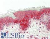
Recognizes a protein of 95 kDa , which is identified as villin. It is a major constituent in the microvilli, which compose the brush border of epithelial cells forming absorptive surfaces of the intestinal and renal proximal tubular epithelia. Anti-Villin labels the brush border area in the gastrointestinal mucosal epithelium and urogenital tract. Among neoplasms, villin is predominantly expressed in tumors of colorectal origin. Antibody to villin is useful in identifying malignant cells from primary and metastatic colorectal carcinomas. This antibody also labels Merkel cells of the skin.
Applications
- IHC - Paraffin (0.5 - 1 µg/ml)
|
Performing IHC? See our complete line of Immunohistochemistry Reagents including antigen retrieval solutions, blocking agents
ABC Detection Kits and polymers, biotinylated secondary antibodies, substrates and more.
|
Usage
Immunohistology (Formalin-fixed - 0.25-0.5 ug/ml for 30 minutes at RT - Staining of formalin-fixed tissues requires boiling tissue sections in 10 mM citrate buffer, pH 6.0, for 10-20 min followed by cooling at RT for 20 minutes.) Optimal dilution for a specific application should be determined.
Presentation
10 mM PBS
Storage
Store at -20°C to -80°C for up to 2 years.
Restrictions
For research use only. Intended for use by laboratory professionals.
About VIL1 / Villin
Publications (0)
Customer Reviews (0)
Featured Products
Species:
Human, Mouse
Applications:
ICC, Immunofluorescence, Western blot
Species:
Human, Mouse, Rat
Applications:
IHC, IHC - Paraffin, Western blot
Species:
Human, Mouse
Applications:
IHC, IHC - Paraffin, Western blot
Species:
Human, Mouse, Pig
Applications:
Western blot
Species:
Human, Mouse
Applications:
IHC, IHC - Paraffin, Western blot, Flow Cytometry
Request SDS/MSDS
To request an SDS/MSDS form for this product, please contact our Technical Support department at:
Technical.Support@LSBio.com
Requested From: United States
Date Requested: 4/10/2025
Date Requested: 4/10/2025

