Login
Registration enables users to use special features of this website, such as past
order histories, retained contact details for faster checkout, review submissions, and special promotions.
order histories, retained contact details for faster checkout, review submissions, and special promotions.
Forgot password?
Registration enables users to use special features of this website, such as past
order histories, retained contact details for faster checkout, review submissions, and special promotions.
order histories, retained contact details for faster checkout, review submissions, and special promotions.
Quick Order
Products
Antibodies
ELISA and Assay Kits
Research Areas
Infectious Disease
Resources
Purchasing
Reference Material
Contact Us
Location
Corporate Headquarters
Vector Laboratories, Inc.
6737 Mowry Ave
Newark, CA 94560
United States
Telephone Numbers
Customer Service: (800) 227-6666 / (650) 697-3600
Contact Us
Additional Contact Details
Login
Registration enables users to use special features of this website, such as past
order histories, retained contact details for faster checkout, review submissions, and special promotions.
order histories, retained contact details for faster checkout, review submissions, and special promotions.
Forgot password?
Registration enables users to use special features of this website, such as past
order histories, retained contact details for faster checkout, review submissions, and special promotions.
order histories, retained contact details for faster checkout, review submissions, and special promotions.
Quick Order
| Catalog Number | Size | Price |
|---|---|---|
| LS-C104402-50 | 50 µg | $356 |
| LS-C104402-100 | 100 µg | $396 |


1 of 3
2 of 3
3 of 3
Polyclonal Rabbit anti‑Mouse CXCL10 / IP‑10 Antibody (WB) LS‑C104402
Polyclonal Rabbit anti‑Mouse CXCL10 / IP‑10 Antibody (WB) LS‑C104402
Note: This antibody replaces LS-C54154, LS-C16064
Antibody:
CXCL10 / IP-10 Rabbit anti-Mouse Polyclonal Antibody
Application:
WB, ELISA
Reactivity:
Mouse
Format:
Unconjugated, Unmodified
Other formats:
Toll Free North America
 (800) 227-6666
For Research Use Only
Overview
Antibody:
CXCL10 / IP-10 Rabbit anti-Mouse Polyclonal Antibody
Application:
WB, ELISA
Reactivity:
Mouse
Format:
Unconjugated, Unmodified
Other formats:
Specifications
Description
IP-10 antibody LS-C104402 is an unconjugated rabbit polyclonal antibody to mouse IP-10 (CXCL10). Validated for ELISA and WB. Cited in 2 publications.
Target
Mouse CXCL10 / IP-10
Synonyms
CXCL10 | C-X-C motif chemokine 10 | Gamma-IP10 | IP-10 | Gamma IP10 | GIP-10 | Mob-1 | Small-inducible cytokine B10 | Crg-2 | IFI10 | INP10 | SCYB10
Host
Rabbit
Reactivity
Mouse
(tested or 100% immunogen sequence identity)
Clonality
Polyclonal
Conjugations
Unconjugated.
Also available conjugated with Biotin.
Purification
Immunoaffinity purified
Modifications
Unmodified
Immunogen
E.coli derived recombinant Murine IP-0.
Specificity
Mouse IP-10
Applications
- Western blot (0.1 - 0.2 µg/ml)
- ELISA (0.5 - 2 µg/ml)
Usage
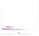
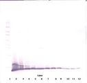
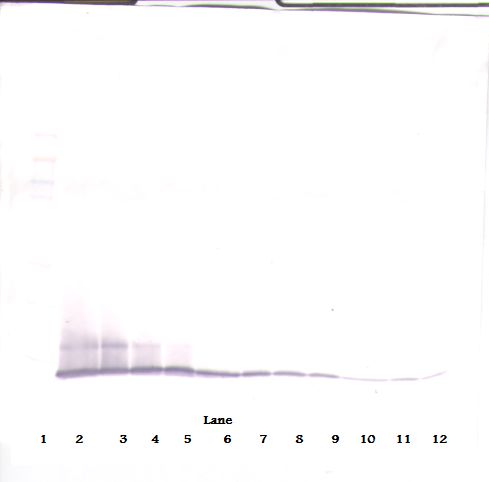
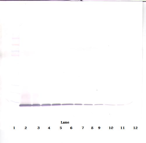
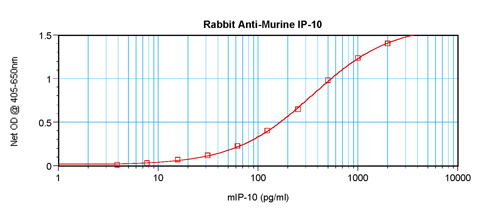
Immunohistochemistry: This antibody stained colchicine injected mouse brain (including the hippocampus region) tissue. The primary antibody was incubated at 1 mg/ml overnight at 4°C. This was followed by a peroxidase conjugated secondary antibody and then a fluorescein Tyramide Signal Amplification (TSA) reagent. Optimal concentrations and conditions may vary. Neutralization: To yield one-half maximal inhibition [ND] of the biological activity of mIP-10 (100 ng/ml), a concentration of 10 ug/ml of this antibody is required. ELISA: To detect mIP-10 by sandwich ELISA (using 100 ul/well antibody solution) a concentration of 0.5-2 ug/ml of this antibody is required. This antigen affinity purified antibody, in conjunction with Biotinylated Anti-Murine IP-10 (LS-C104391) as a detection antibody, allows the detection of at least 0.2-0.4 ng/well of recombinant mIP-10. Western Blot: To detect mIP-10 by Western Blot analysis this antibody can be used at a concentration of 0.1-0.2 ug/ml. Used in conjunction with compatible secondary reagents the detection limit for recombinant mIP-10 is 1.5-3 ng/lane, under either reducing or non-reducing conditions.
Presentation
Lyophilized from PBS, pH 7.2
Reconstitution
sterile ddH20
Storage
Store Lyophilized at room temperature up to 1 month; Reconstituted for up to 2 weeks at 2°C to 8°C. Aliquot and freeze at -20°C for long term storage. Avoid freeze/thaw cycles.
Restrictions
For research use only. Intended for use by laboratory professionals.
About CXCL10 / IP-10
LSBio Ratings
CXCL10 / IP-10 Antibody for WB/Western, ELISA LS-C104402 has an LSBio Rating of
Publications (4.1)
Learn more about The LSBio Ratings Algorithm
Publications (2)
Cisplatin Facilitates Radiation-Induced Abscopal Effects in Conjunction with PD-1 Checkpoint Blockade Through CXCR3/CXCL10-Mediated T-cell Recruitment. Ren Luo, Elke Firat, Simone Gaedicke, Elena Guffart, Tsubasa Watanabe, Gabriele Niedermann. Clinical cancer research : an official journal of the American Association for Cancer Research. 2019 December;25:7243-7255.
Long non-coding RNA LINC00472 inhibits oral squamous cell carcinoma via miR-4311/GNG7 axis. Chen Zou, Xiaozhi Lv, Haigang Wei, Siyuan Wu, Jing Song, Zhe Tang, Shiwei Liu, Xia Li, Yilong Ai. Bioengineered. 2022 March;13:6371-6382.
Customer Reviews (0)
Featured Products
Species:
Human
Applications:
IHC, IHC - Paraffin, Western blot, ELISA
Request SDS/MSDS
To request an SDS/MSDS form for this product, please contact our Technical Support department at:
Technical.Support@LSBio.com
Requested From: United States
Date Requested: 4/15/2025
Date Requested: 4/15/2025


