Login
Registration enables users to use special features of this website, such as past
order histories, retained contact details for faster checkout, review submissions, and special promotions.
order histories, retained contact details for faster checkout, review submissions, and special promotions.
Forgot password?
Registration enables users to use special features of this website, such as past
order histories, retained contact details for faster checkout, review submissions, and special promotions.
order histories, retained contact details for faster checkout, review submissions, and special promotions.
Quick Order
Products
Antibodies
ELISA and Assay Kits
Research Areas
Infectious Disease
Resources
Purchasing
Reference Material
Contact Us
Location
Corporate Headquarters
Vector Laboratories, Inc.
6737 Mowry Ave
Newark, CA 94560
United States
Telephone Numbers
Customer Service: (800) 227-6666 / (650) 697-3600
Contact Us
Additional Contact Details
Login
Registration enables users to use special features of this website, such as past
order histories, retained contact details for faster checkout, review submissions, and special promotions.
order histories, retained contact details for faster checkout, review submissions, and special promotions.
Forgot password?
Registration enables users to use special features of this website, such as past
order histories, retained contact details for faster checkout, review submissions, and special promotions.
order histories, retained contact details for faster checkout, review submissions, and special promotions.
Quick Order
| Catalog Number | Size | Price |
|---|---|---|
| LS-C389961-7 | 7 ml | $574 |
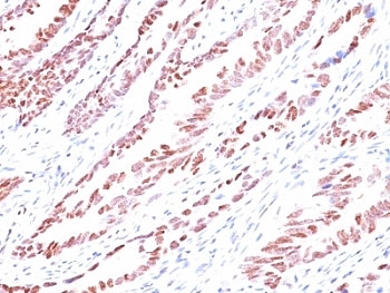
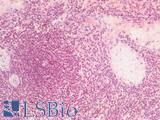
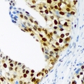
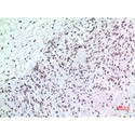
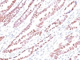
Monoclonal Mouse anti‑Human CDKN1C / p57 Kip2 Antibody (clone KP10, Ready‑to‑Use, IHC, IF) LS‑C389961
Monoclonal Mouse anti‑Human CDKN1C / p57 Kip2 Antibody (clone KP10, Ready‑to‑Use, IHC, IF) LS‑C389961
Antibody:
CDKN1C / p57 Kip2 Mouse anti-Human Monoclonal (Ready-to-Use) (KP10) Antibody
Application:
IHC, IHC-P, IF, Flo
Reactivity:
Human, Mouse
Format:
Unconjugated, Ready-to-Use
Other formats:
Toll Free North America
 (800) 227-6666
For Research Use Only
Overview
Antibody:
CDKN1C / p57 Kip2 Mouse anti-Human Monoclonal (Ready-to-Use) (KP10) Antibody
Application:
IHC, IHC-P, IF, Flo
Reactivity:
Human, Mouse
Format:
Unconjugated, Ready-to-Use
Other formats:
Specifications
Description
P57 Kip2 antibody LS-C389961 is an unconjugated mouse monoclonal antibody to p57 Kip2 (CDKN1C) from human. It is reactive with human and mouse. Validated for Flow, IF and IHC.
Target
Human CDKN1C / p57 Kip2
Synonyms
CDKN1C | BWS | KIP2 | IMAGE | p57Kip2 | p57 | p57 KIP2 | Beckwith-Wiedemann syndrome | WBS | BWCR
Host
Mouse
Reactivity
Human, Mouse
(tested or 100% immunogen sequence identity)
Clonality
IgG2b,k
Monoclonal
Clone
KP10
Conjugations
Unconjugated
Purification
Protein G purified
Modifications
Ready-to-Use.
Also available Unmodified or Azide-free, BSA-free.
Immunogen
Recombinant human protein.
Specificity
Human CDKN1C / p57 Kip2
Applications
- IHC
- IHC - Paraffin (1:1)
- Immunofluorescence
- Flow Cytometry

|
Performing IHC? See our complete line of Immunohistochemistry Reagents including antigen retrieval solutions, blocking agents
ABC Detection Kits and polymers, biotinylated secondary antibodies, substrates and more.
|
Usage
The applications listed have been tested for the unmodified form of this product. Other forms have not been tested. Recognizes a protein of 57kDa, identified as p57Kip2. It shows no cross-reaction with p27Kip1. p57Kip2 is a potent tight-binding inhibitor of several G1 cyclin complexes, and is a negative regulator of cell proliferation. Anti-p57 has been used as an aide in identification of complete hydatidiform mole (CHM) (no nuclear labeling of cytotrophoblasts and stromal cells) from partial hydatidiform mole (PHM) in which both cytotrophoblasts and stromal cells stain. The histological differentiation of complete mole, partial mole, and hydropic spontaneous abortion is problematic. Most complete hydatidiform moles are diploid, whereas most partial moles are triploid. Ploidy studies will identify partial moles, but will not differentiate complete moles from non-molar gestations. Complete moles carry a high risk of persistent disease and choriocarcinoma, while partial moles have a very low risk. In normal placenta, many cytotrophoblast nuclei and stromal cells are labeled with this antibody. Similar findings apply to PHM and hydropic abortus tissues. Intervillous trophoblastic islands (IVTIs) demonstrate nuclear labeling in all three entities and serve as an internal control. Optimal dilution of the p57 antibody to be determined by the researcher. Staining of formalin-fixed tissues requires boiling tissue sections in 10mM Citrate buffer, pH 6.0, for 10-20 min followed by cooling at RT for 20 minutes. The prediluted format is supplied in a dropper bottle and is optimized for use in IHC. After epitope retrieval step (if required), drip mAb solution onto the tissue section and incubate at RT for 30 min.
Presentation
1X PBS, 0.1 mg/ml BSA, 0.05% sodium azide
Storage
Store at 2°C to 8°C.
Restrictions
For research use only. Intended for use by laboratory professionals.
About CDKN1C / p57 Kip2
Publications (0)
Customer Reviews (0)
Featured Products
Species:
Human
Applications:
IHC, IHC - Paraffin, Immunofluorescence, Western blot, ELISA
Species:
Human, Mouse
Applications:
IHC, IHC - Paraffin, Western blot, Immunoprecipitation
Species:
Human, Mouse
Applications:
IHC, ICC, Immunofluorescence, Western blot
Species:
Human, Mouse, Rat
Applications:
IHC, Western blot, ELISA
Species:
Human, Mouse
Applications:
IHC, IHC - Paraffin, Immunofluorescence, Western blot, Flow Cytometry

Species:
Human, Mouse
Applications:
IHC, IHC - Paraffin, Immunofluorescence, Western blot, Flow Cytometry
Request SDS/MSDS
To request an SDS/MSDS form for this product, please contact our Technical Support department at:
Technical.Support@LSBio.com
Requested From: United States
Date Requested: 2/5/2025
Date Requested: 2/5/2025


