Login
Registration enables users to use special features of this website, such as past
order histories, retained contact details for faster checkout, review submissions, and special promotions.
order histories, retained contact details for faster checkout, review submissions, and special promotions.
Forgot password?
Registration enables users to use special features of this website, such as past
order histories, retained contact details for faster checkout, review submissions, and special promotions.
order histories, retained contact details for faster checkout, review submissions, and special promotions.
Quick Order
Products
Antibodies
ELISA and Assay Kits
Research Areas
Infectious Disease
Resources
Purchasing
Reference Material
Contact Us
Location
Corporate Headquarters
Vector Laboratories, Inc.
6737 Mowry Ave
Newark, CA 94560
United States
Telephone Numbers
Customer Service: (800) 227-6666 / (650) 697-3600
Contact Us
Additional Contact Details
Login
Registration enables users to use special features of this website, such as past
order histories, retained contact details for faster checkout, review submissions, and special promotions.
order histories, retained contact details for faster checkout, review submissions, and special promotions.
Forgot password?
Registration enables users to use special features of this website, such as past
order histories, retained contact details for faster checkout, review submissions, and special promotions.
order histories, retained contact details for faster checkout, review submissions, and special promotions.
Quick Order
| Catalog Number | Size | Price |
|---|---|---|
| LS-C756577-100 | 100 µg | $470 |
1 of 11
2 of 11
3 of 11
4 of 11
5 of 11
6 of 11
7 of 11
8 of 11
9 of 11
10 of 11
11 of 11
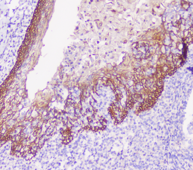
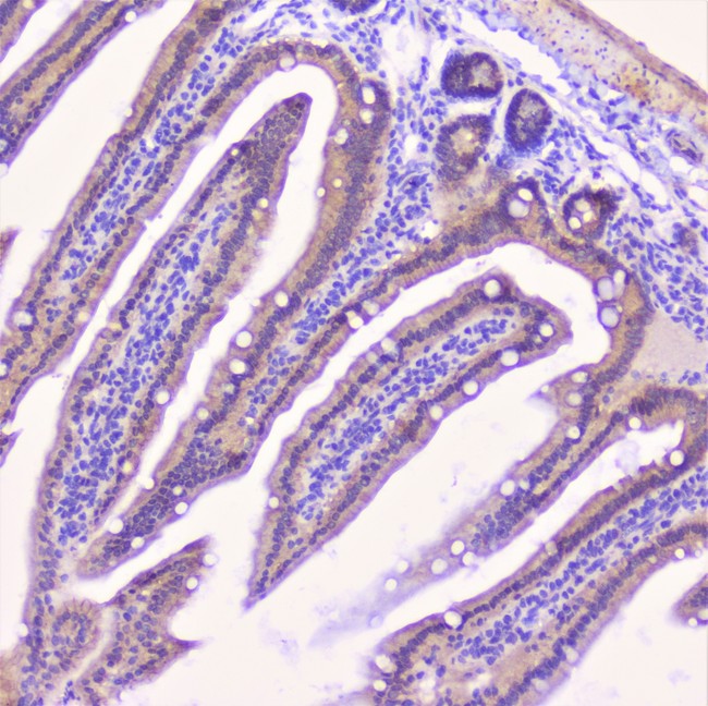
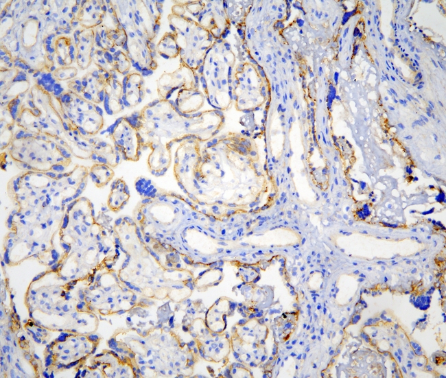
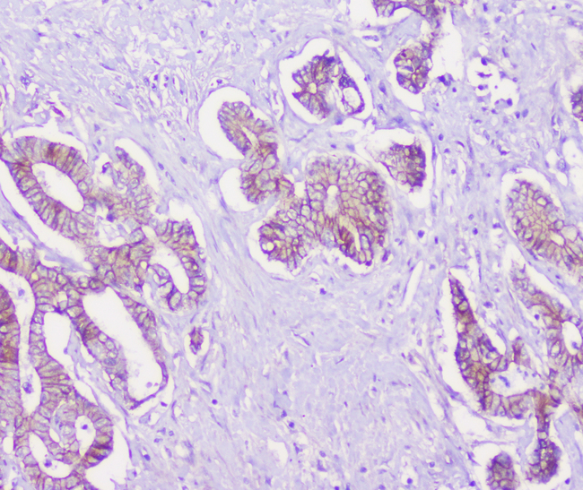
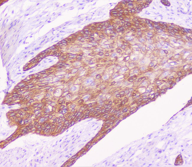
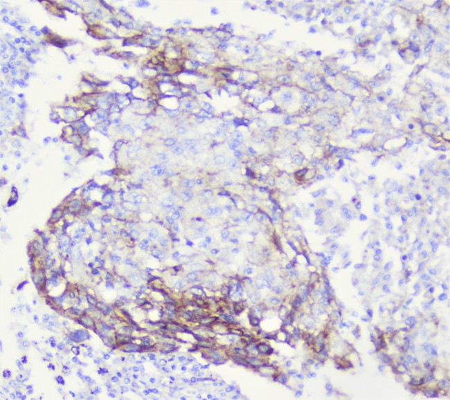
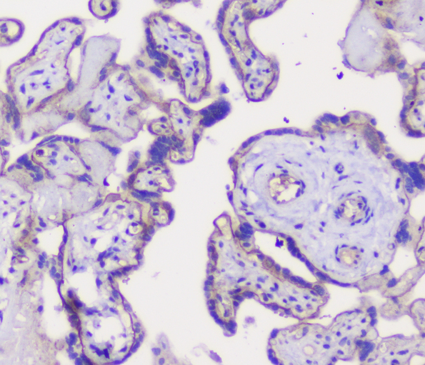
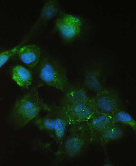
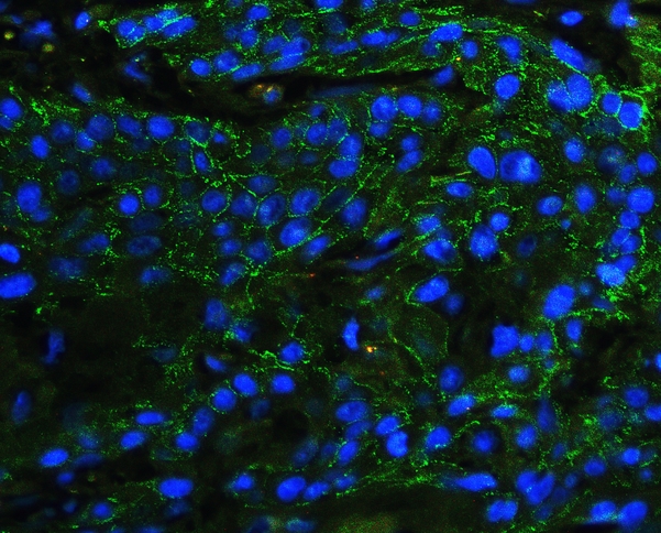
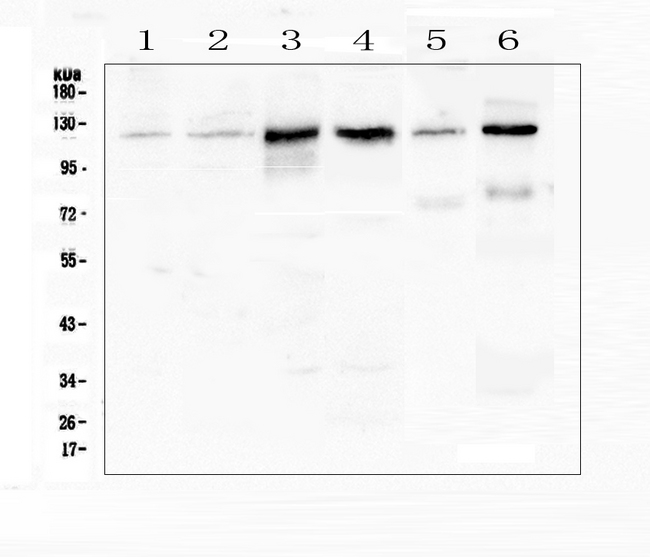
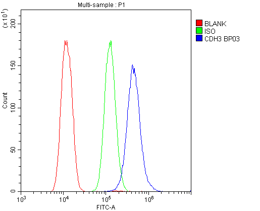
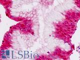
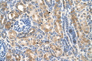
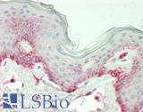
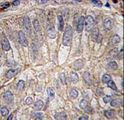
Polyclonal Rabbit anti‑Human CDH3 / P‑Cadherin Antibody (IHC, WB) LS‑C756577
Polyclonal Rabbit anti‑Human CDH3 / P‑Cadherin Antibody (IHC, WB) LS‑C756577
Antibody:
CDH3 / P-Cadherin Rabbit anti-Human Polyclonal Antibody
Application:
IHC-P, WB, ELISA
Reactivity:
Human, Mouse, Rat
Format:
Unconjugated, Unmodified
Toll Free North America
 (800) 227-6666
For Research Use Only
Overview
Antibody:
CDH3 / P-Cadherin Rabbit anti-Human Polyclonal Antibody
Application:
IHC-P, WB, ELISA
Reactivity:
Human, Mouse, Rat
Format:
Unconjugated, Unmodified
Specifications
Description
P-Cadherin antibody LS-C756577 is an unconjugated rabbit polyclonal antibody to P-Cadherin (CDH3) from human. It is reactive with human, mouse and rat. Validated for ELISA, IHC and WB.
Target
Human CDH3 / P-Cadherin
Synonyms
CDH3 | Cadherin-3 | HJMD | P-cadherin | CDHP | PCAD | Placental cadherin
Host
Rabbit
Reactivity
Human, Mouse, Rat
(tested or 100% immunogen sequence identity)
Clonality
IgG
Polyclonal
Conjugations
Unconjugated
Purification
Immunogen affinity purified
Modifications
Unmodified
Immunogen
E. coli-derived human P cadherin recombinant protein (Position: Q126-H336).
Specificity
No cross reactivity with other proteins.
Applications
- IHC - Paraffin (0.5 - 1 µg/ml)
- Western blot (0.1 - 0.5 µg/ml)
- ELISA (0.1 - 0.5 µg/ml)

|
Performing IHC? See our complete line of Immunohistochemistry Reagents including antigen retrieval solutions, blocking agents
ABC Detection Kits and polymers, biotinylated secondary antibodies, substrates and more.
|
Usage
Applications should be user optimized.
Presentation
Lyophilized from 0.2mg Na2HPO4, 0.9mg NaCl, 0.05mg sodium azide, 4mg Trehalose
Reconstitution
Add 0.2ml of distilled water will yield a concentration of 500µg/ml.
Storage
After reconstitution, may be stored at 4°C for 1 month. For long-term storage and to avoid freeze-thaw cycles, aliquot and store at -20°C.
Restrictions
For research use only. Intended for use by laboratory professionals.
About CDH3 / P-Cadherin
Publications (0)
Customer Reviews (0)
Featured Products
Species:
Mouse
Applications:
IHC, Western blot, ELISA, Functional Assay
Species:
Human, Mouse
Applications:
IHC, IHC - Paraffin, Western blot, Flow Cytometry
Species:
Human
Applications:
IHC, IHC - Paraffin, Immunofluorescence, Western blot, Immunoprecipitation, ELISA, Functional Assay
Species:
Human
Applications:
IHC, IHC - Paraffin, Western blot
Species:
Human
Applications:
IHC, IHC - Paraffin, Immunofluorescence, Western blot
Species:
Human
Applications:
IHC, IHC - Paraffin, Western blot
Request SDS/MSDS
To request an SDS/MSDS form for this product, please contact our Technical Support department at:
Technical.Support@LSBio.com
Requested From: United States
Date Requested: 4/18/2025
Date Requested: 4/18/2025


