Login
Registration enables users to use special features of this website, such as past
order histories, retained contact details for faster checkout, review submissions, and special promotions.
order histories, retained contact details for faster checkout, review submissions, and special promotions.
Forgot password?
Registration enables users to use special features of this website, such as past
order histories, retained contact details for faster checkout, review submissions, and special promotions.
order histories, retained contact details for faster checkout, review submissions, and special promotions.
Quick Order
Products
Antibodies
ELISA and Assay Kits
Research Areas
Infectious Disease
Resources
Purchasing
Reference Material
Contact Us
Location
Corporate Headquarters
Vector Laboratories, Inc.
6737 Mowry Ave
Newark, CA 94560
United States
Telephone Numbers
Customer Service: (800) 227-6666 / (650) 697-3600
Contact Us
Additional Contact Details
Login
Registration enables users to use special features of this website, such as past
order histories, retained contact details for faster checkout, review submissions, and special promotions.
order histories, retained contact details for faster checkout, review submissions, and special promotions.
Forgot password?
Registration enables users to use special features of this website, such as past
order histories, retained contact details for faster checkout, review submissions, and special promotions.
order histories, retained contact details for faster checkout, review submissions, and special promotions.
Quick Order
| Catalog Number | Size | Price |
|---|---|---|
| LS-C800179-100 | 100 µg | $539 |
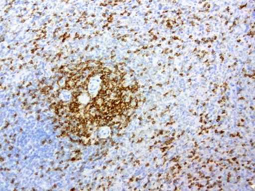
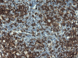
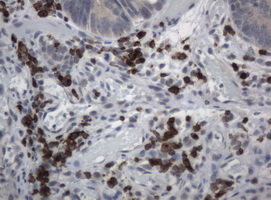
![CD2 Antibody - IHC staining of paraffin-embedded human tonsil using anti-CD2 clone UMAB6 mouse monoclonal antibody at 1:200 of 0.6mg/mL and detection with Polink2 Broad HRP DAB.requires heat-induced epitope retrieval with Accel pH8.7 at 95-100C 30 minutes [do not let boil] or 10 min in pressure cooker. The image shows strong membranous and cytoplasmic staining in >50 % of non germinal center cells of tonsil and <20% of the germinal center cells. No staining was seen in the squamous epithelia cells.](https://lsbio-7d62.kxcdn.com/image2/cd2-antibody-clone-umab6-carrier-free-ls-c800179/549082_4580629.jpg)
1 of 15
2 of 15
3 of 15
4 of 15
5 of 15
6 of 15
7 of 15
8 of 15
9 of 15
10 of 15
11 of 15
12 of 15
13 of 15
14 of 15
15 of 15
Monoclonal Mouse anti‑Human CD2 Antibody (clone UMAB6, Carrier‑free, IHC, IF, WB) LS‑C800179
Monoclonal Mouse anti‑Human CD2 Antibody (clone UMAB6, Carrier‑free, IHC, IF, WB) LS‑C800179
Antibody:
CD2 Mouse anti-Human Monoclonal (Carrier-free) (UMAB6) Antibody
Application:
IHC, IF, WB, Flo, PMA
Reactivity:
Human
Format:
Unconjugated, Carrier-free
Other formats:
Toll Free North America
 (800) 227-6666
For Research Use Only
Overview
Antibody:
CD2 Mouse anti-Human Monoclonal (Carrier-free) (UMAB6) Antibody
Application:
IHC, IF, WB, Flo, PMA
Reactivity:
Human
Format:
Unconjugated, Carrier-free
Other formats:
Specifications
Description
CD2 antibody LS-C800179 is an unconjugated mouse monoclonal antibody to human CD2. Validated for Flow, IF, IHC, PMA and WB.
Target
Human CD2
Synonyms
CD2 | CD2 antigen | Erythrocyte receptor | LFA-3 receptor | LFA-2 | Lymphocyte-function antigen-2 | T11 | SRBC | CD2 molecule | Rosette receptor | T-cell surface antigen CD2
Host
Mouse
Reactivity
Human
(tested or 100% immunogen sequence identity)
Clonality
IgG1
Monoclonal
Clone
UMAB6
Conjugations
Unconjugated
Purification
Affinity chromatography
Modifications
Carrier-free.
Also available Unmodified.
Immunogen
Protein expressed in 293T cell transfected with human CD2 expression vector.
Specificity
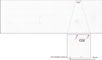
The specificity of this antibody has been validated by testing against a high-density Protein Microarray containing more than 17,000 recombinant proteins.
Applications
- IHC
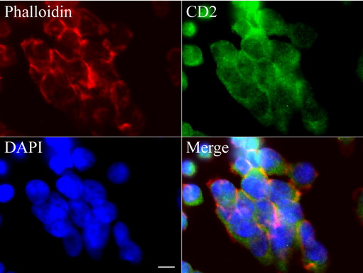
- Immunofluorescence
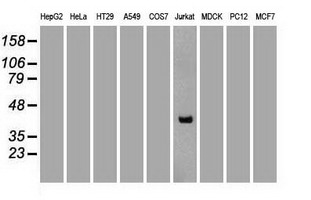
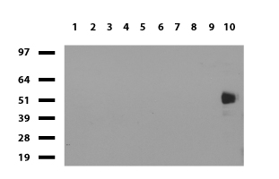
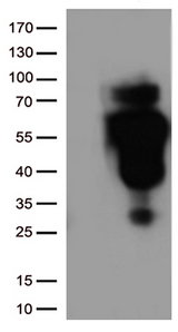
- Western blot (1:2000)
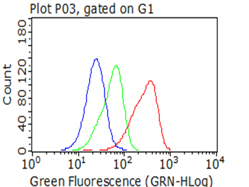
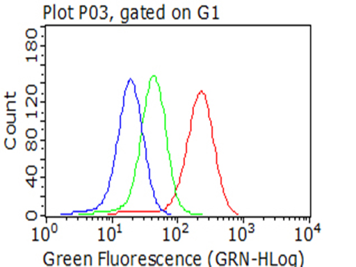
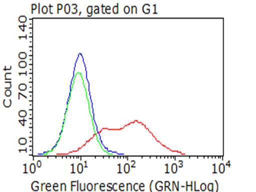
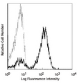
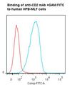
- Flow Cytometry
- Protein Microarray

|
Performing IHC? See our complete line of Immunohistochemistry Reagents including antigen retrieval solutions, blocking agents
ABC Detection Kits and polymers, biotinylated secondary antibodies, substrates and more.
|
Usage
Applications should be user optimized.
Presentation
PBS, pH 7.3, 8% Trehalose
Reconstitution
Reconstitute with PBS pH 7.3. To use this carrier-free antibody for conjugation experiment, we strongly recommend you to perform another round of desalting process.
Storage
Store at -20°C. Avoid freeze-thaw cycles.
Restrictions
For research use only. Intended for use by laboratory professionals.
About CD2
Publications (0)
Customer Reviews (0)
Featured Products
Species:
Human, Baboon, Monkey, Pig
Applications:
IHC, IHC - Frozen, Flow Cytometry
Species:
Mouse
Applications:
IHC, IHC - Frozen, Flow Cytometry
Species:
Rat
Applications:
IHC, IHC - Paraffin, IHC - Frozen, Flow Cytometry
Species:
Human
Applications:
IHC, IHC - Frozen, Immunofluorescence, Immunoprecipitation, Flow Cytometry, ELISA, Functional Assay
Species:
Human
Applications:
Western blot, Flow Cytometry, Functional Assay
Request SDS/MSDS
To request an SDS/MSDS form for this product, please contact our Technical Support department at:
Technical.Support@LSBio.com
Requested From: United States
Date Requested: 4/16/2025
Date Requested: 4/16/2025


