Products
Research Areas
COVID-19
Resources
Login
Quick Order
Cart Cart lightblue
Login
Registration enables users to use special features of this website, such as past
order histories, retained contact details for faster checkout, review submissions, and special promotions.


Fields marked with a * are required.

Login
Quick Order
Contact Us

Location


Corporate Headquarters

Vector Laboratories, Inc.
6737 Mowry Ave
Newark, CA 94560
United States

Telephone Numbers


Customer Service: (800) 227-6666 / (650) 697-3600


Contact Us


Additional Contact Details

Login
Registration enables users to use special features of this website, such as past
order histories, retained contact details for faster checkout, review submissions, and special promotions.


Fields marked with a * are required.

Login
Quick Order
Catalog Number Size Price
LS-C343990-10 10 µg $318 
LS-C343990-100 100 µg (0.5 mg/ml) $470 
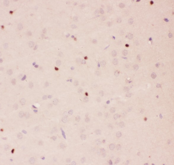
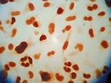
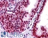
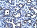
APEX1 / APE1 Antibody - APE1 antibody IHC-paraffin: Rat Brain Tissue.
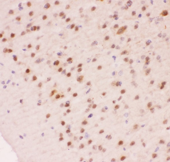
APEX1 / APE1 Antibody - APE1 antibody IHC-paraffin: Mouse Brain Tissue.
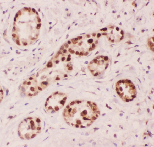
APEX1 / APE1 Antibody - APE1 antibody IHC-paraffin: Human Lung Cancer Tissue.
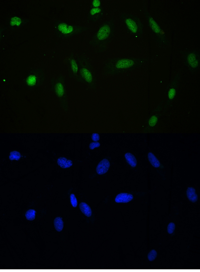
APEX1 / APE1 Antibody - IF analysis of APEX1 using anti-APEX1 antibody APEX1 was detected in immunocytochemical section of U20S cell. Enzyme antigen retrieval was performed using IHC enzyme antigen retrieval reagent for 15 mins. The tissue section was blocked with 10% goat serum. The tissue section was then incubated with 2µg/mL rabbit anti-APEX1 Antibody overnight at 4°C. DyLight®488 Conjugated Goat Anti-Rabbit IgG was used as secondary antibody at 1:100 dilution and incubated for 30 minutes at 37°C. The section was counterstained with DAPI. Visualize using a fluorescence microscope and filter sets appropriate for the label used.
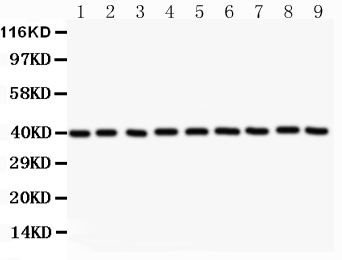
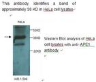
APEX1 / APE1 Antibody - Western blot analysis of APEX1 using anti-APEX1 antibody. Electrophoresis was performed on a 5-20% SDS-PAGE gel at 70V (Stacking gel) / 90V (Resolving gel) for 2-3 hours. The sample well of each lane was loaded with 50ug of sample under reducing conditions. Lane 1: NRK Whole Cell Lysate Lane 2: HELA Whole Cell Lysate Lane 3: PC-12 Whole Cell Lysate Lane 4: RH35 Whole Cell Lysate Lane 5: HEPA Whole Cell Lysate Lane 6: MCF Whole Cell Lysate Lane 7: A549 Whole Cell Lysate Lane 8: Human Placenta Tissue Lysate Lane 9: A431 Whole Cell Lysate After Electrophoresis, proteins were transferred to a Nitrocellulose membrane at 150mA for 50-90 minutes. Blocked the membrane with 5% Non-fat Milk/ TBS for 1.5 hour at RT. The membrane was incubated with rabbit anti-APEX1 antigen affinity purified polyclonal antibody at 0.5 µg/mL overnight at 4°C, then washed with TBS-0.1% Tween 3 times with 5 minutes each and probed with a goat anti-rabbit IgG-HRP secondary antibody at a dilution of 1:10000 for 1.5 hour at RT. The signal is developed using an Enhanced Chemiluminescent detection (ECL) kit with Tanon 5200 system. A specific band was detected for APEX1 at approximately 39KD. The expected band size for APEX1 is at 39KD.
APEX1 / APE1 Antibody - APE1 antibody Western blot. All lanes: Anti APEX1 at 0.5 ug/ml. WB: Recombinant Human APEX1 Protein 0.5ng. Predicted band size: 45 kD. Observed band size: 45 kD.
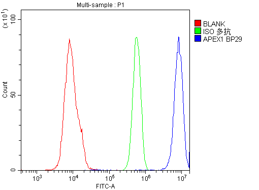
APEX1 / APE1 Antibody - Flow Cytometry analysis of U937 cells using anti-APEX1 antibody. Overlay histogram showing U937 cells stained with anti-APEX1 antibody (Blue line). The cells were blocked with 10% normal goat serum. And then incubated with rabbit anti-APEX1 Antibody (1µg/10E6 cells) for 30 min at 20°C. DyLight®488 conjugated goat anti-rabbit IgG (5-10µg/10E6 cells) was used as secondary antibody for 30 minutes at 20°C. Isotype control antibody (Green line) was rabbit IgG (1µg/10E6 cells) used under the same conditions. Unlabelled sample (Red line) was also used as a control.
1 of 7
2 of 7
3 of 7
4 of 7
5 of 7
6 of 7
7 of 7

Polyclonal Rabbit anti‑Human APEX1 / APE1 Antibody (aa2‑318, IHC, WB) LS‑C343990

Polyclonal Rabbit anti‑Human APEX1 / APE1 Antibody (aa2‑318, IHC, WB) LS‑C343990

Note: This antibody replaces LS-C387982
Antibody:
APEX1 / APE1 Rabbit anti-Human Polyclonal (aa2-318) Antibody
Application:
IHC, IHC-P, WB
Reactivity:
Human, Mouse, Rat
Format:
Unconjugated, Unmodified
Price
Catalog Number
$318
LS-C343990-10
Toll Free North America
(800) 227-6666
For Research Use Only

Overview

Antibody:
APEX1 / APE1 Rabbit anti-Human Polyclonal (aa2-318) Antibody
Application:
IHC, IHC-P, WB
Reactivity:
Human, Mouse, Rat
Format:
Unconjugated, Unmodified

Specifications

Description
APE1 antibody LS-C343990 is an unconjugated rabbit polyclonal antibody to APE1 (APEX1) (aa2-318) from human. It is reactive with human, mouse and rat. Validated for IHC and WB.
Target
Human APEX1 / APE1
Synonyms
APEX1 | AP endonuclease class I | AP lyase | APE | APE-1 | APEN | APE1 | APEX nuclease | Redox factor-1 | AP endonuclease 1 | APEX | APX | Protein REF-1 | REF-1 | REF1
Host
Rabbit
Reactivity
Human, Mouse, Rat (tested or 100% immunogen sequence identity)
Clonality
Polyclonal
Conjugations
Unconjugated
Purification
Immunogen affinity purified
Modifications
Unmodified
Immunogen
E.coli-derived human APE1 recombinant protein (Position: P2-L318). Human APE1 shares 94% and 93% amino acid (aa) sequences identity with mouse and rat APE1, respectively.
Epitope
aa2-318
Applications
  • IHC
  • IHC - Paraffin
  • Western blot
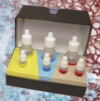
Performing IHC? See our complete line of Immunohistochemistry Reagents including antigen retrieval solutions, blocking agents ABC Detection Kits and polymers, biotinylated secondary antibodies, substrates and more.
Presentation
Lyophilized from 0.2mg Na2HPO4, 5mg BSA, 0.9mg NaCl, 0.05mg sodium azide.
Reconstitution
Add 0.2ml of distilled water will yield a concentration of 500µg/ml.
Storage
At -20°C for 1 year. After reconstitution, at 4°C for 1 month. It can also be aliquotted and stored frozen at -20°C for a longer time. Avoid freeze-thaw cycles.
Restrictions
For research use only. Intended for use by laboratory professionals.
Guarantee
This antibody carries the LSBio 100% Guarantee.
LSBio Guarantee
About APEX1 / APE1
P27695 NM_001641 NP_001632.2

Publications (0)

Customer Reviews (0)


Request SDS/MSDS

To request an SDS/MSDS form for this product, please contact our Technical Support department at:

Technical.Support@LSBio.com

Requested From: United States
Date Requested: 1/4/2025