Products
Research Areas
COVID-19
Resources
Login
Quick Order
Cart Cart lightblue
Login
Registration enables users to use special features of this website, such as past
order histories, retained contact details for faster checkout, review submissions, and special promotions.


Fields marked with a * are required.

Login
Quick Order
Contact Us

Location


Corporate Headquarters

Vector Laboratories, Inc.
6737 Mowry Ave
Newark, CA 94560
United States

Telephone Numbers


Customer Service: (800) 227-6666 / (650) 697-3600


Contact Us


Additional Contact Details

Login
Registration enables users to use special features of this website, such as past
order histories, retained contact details for faster checkout, review submissions, and special promotions.


Fields marked with a * are required.

Login
Quick Order
Catalog Number Size Price
LS-C744816-25 25 µl (1 mg/ml) $304 
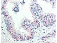
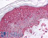
AKT1 Antibody - Immunohistochemistry of Mouse anti-AKT pS473 antibody. Tissue: human prostate tissue. Fixation: formalin fixed paraffin embedded. Antigen retrieval: not required. Primary antibody: AKT pS473 antibody at 20 µg/mL for 1 h at RT. Secondary antibody: Dako's Techmate streptavidin-biotin reagents at 1:10,000 for 45 min at RT. Localization: AKT pS473 is nuclear and occasionally cytoplasmic. Staining: AKT pS473 as precipitated red signal with hematoxylin purple nuclear counterstain.
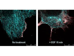
AKT1 Antibody - Immunofluorescence confocal microscopy of Mouse Anti-AKT pS473 antibody. Tissue: EGF treated A431 cells. Fixation: 0.5% PFA. Antigen retrieval: EGF 15 min. Primary antibody: AKT pS473 antibody at 10 µg/mL for 1 h at RT. Secondary antibody: DyLight 488 Goat anti-Rabbit IgG, MAb anti-AKT pS473, atto-647N anti-Mouse IgG (Active Motif). at 1:10,000 for 45 min at RT. Localization: AKT pS473 is nuclear and occasionally cytoplasmic. Staining: AKT pS473 as red signal with tubulin (cyan).
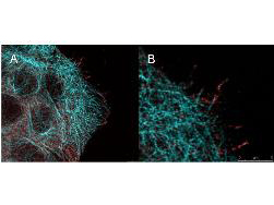
AKT1 Antibody - High resolution STED immunofluorescence nanoscopy of Mouse anti-AKT pS473 antibody. Tissue: A431 cells. The merge images (A) and at high magnification (B) show phosphorylated AKT colocalized with the distal microtubules. Fixation: 4% paraformaldehyde for 5 min and after washes blocked with 10% NGS/0.2% Triton X-100 for 30 min. Antigen retrieval: serum deprivation for 12 h. Primary antibody: AKT pS473 antibody at 10 µg/mL and a-tubulin (cyan) at 1.4 µg/mL for 1 h at RT. Secondary antibody: Atto 647N anti-Mouse IgG (ATTO TEC GmbH), and DyLight 488 anti-Rabbit IgG were used at 1.0 µg/mL for 1h at RT for indirect detection. Localization: AKT pS473 is in the cytoplasm and also organized at the periphery of the cell. Staining: AKT pS473 as red signal with bis-benzimide (blue) nuclear counterstain.
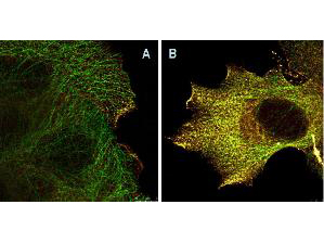
AKT1 Antibody - Immunofluorescence Microscopy of Mouse Anti-AKTpS473 antibody using STED nanoscopy to evaluate AKT activation and migration. Tissue: A431 cells. Antigen retrieval: Panel A: serum starved,unstimulated cells. Panel B: serum starved, EGF stimulated for 15 mins. A massive increase in AKT-pS473 activation, as measured by intensity signal, peaked at 15 minutes and was associated with depolymerized tubulin. Staining: Panel A shows STED data (AKT-pS473, red channel) collected simultaneously with confocal signal (a-tubulin, green channel). Upon stimulation of cells with EGF, a rapid activation of AKT is observed (Panel B) along with a coincident change in the tubulin organization (yellow signal), as well as an extensive cell shape-change (cell membrane folding) and accumulation of AKTpS473 at the cell periphery.
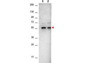
AKT1 Antibody - Western Blot of Mouse Anti-AKTpS473 antibody. Lane 1: A431 cells. Lane 2: A431 cells stimulated for 15 min with EGF. Load: 35 µg per lane. Primary antibody: AKTpS473 antibody at 1:400 for overnight at 4°C. Secondary antibody: DyLight649 Conjugated Anti-AKT pS473 Monoclonal Antibody p/n 200-343-268 at 1:10,000 for 45 min at RT. Block: Blocking Buffer for Fluorescent Western Blotting p/n MB-070 overnight at 4°C. Predicted/Observed size: 56kDa. Other band(s): none.
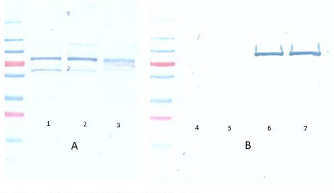
AKT1 Antibody - Western Blot of Mouse Anti-Akt pS473 antibody. A Lane 1) PDGF stimulated NIH 3T3 cells 10 ? Lane 2) NIH 3T3 cells 10 ? Lane 3) Hela whole cell lysate 10 ? (weak signal) B Lane 4) GST negative control protein 100 ng Lane 5) GST negative control protein 25 ng Lane 6) AKT 1 recombinant protein 100 ng Lane 7) AKT 1 recombinant protein 25 ng Block: 5% BSA overnight at 4?. Primary antibody: Monoclonal anti AKT antibody, used at 1:1000 for overnight at 4?. Secondary antibody: HRP Conjugated goat anti mouse lot 20121 1:25K for 45 min at RT. Detection : TMBM-100 for 20 minutes, rinsed with deionized water, dried and scanned on conventional flatbed scanner
AKT1 Antibody - Western Blot of Mouse Anti-AKT pS473 antibody. Lane 1: non-phosphorylated AKT in untreated cells. Lane 2: phosphorylated AKT (indicated by arrowhead at ~56 kDa) on PDGF stimulated NIH/3T3 cell lysates. Load: 10 µg per lane. Primary antibody: AKT pS473 antibody at 1:10,000 in TBS with 0.05% Tween-20 with 1% BSA, for 1 h at 4° C. Secondary antibody: HRP conjugated Gt-a-Mouse IgG was used at a 1:20,000 dilution for 1 h at 4° C with FemtoMax enhanced chemiluminescent reagent Other band(s): none.
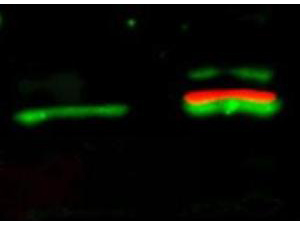
AKT1 Antibody - Western Blot of Mouse Anti-Akt pS473 antibody. Lane 1: unstimulated NIH/3T3 lysates contain inactive unphosphorylated Akt1, green band. Lane 2: PDGF stimulated NIH/3T3 lysate contains both inactive (green band) and activated phosphorylated Akt1 (red band). Load: 10 µg per lane. Primary antibody: rabbit anti-Akt (pan) and mouse anti-Akt pS473 specific antibodies at 1:400 for overnight at 4°C. Secondary antibody: DyLight 549 conjugated anti-rabbit IgG (green) and DyLight 649 conjugated anti-mouse IgG (red) secondary antibodies at 1:10,000 for 45 min at RT. Block: 5% BLOTTO overnight at 4°C.
1 of 8
2 of 8
3 of 8
4 of 8
5 of 8
6 of 8
7 of 8
8 of 8

Monoclonal Mouse anti‑Human AKT1 Antibody (clone 17F6.B11, phospho‑Ser473, IHC, IF, WB) LS‑C744816

Monoclonal Mouse anti‑Human AKT1 Antibody (clone 17F6.B11, phospho‑Ser473, IHC, IF, WB) LS‑C744816

Antibody:
AKT1 Mouse anti-Human Monoclonal (pSer473) (17F6.B11) Antibody
Application:
IHC, IF, WB, IP, Flo, ELISA
Reactivity:
Human, Monkey, Mouse, Rat
Format:
Unconjugated, Unmodified
Price
Catalog Number
$304
LS-C744816-25
Toll Free North America
(800) 227-6666
For Research Use Only

Overview

Antibody:
AKT1 Mouse anti-Human Monoclonal (pSer473) (17F6.B11) Antibody
Application:
IHC, IF, WB, IP, Flo, ELISA
Reactivity:
Human, Monkey, Mouse, Rat
Format:
Unconjugated, Unmodified

Specifications

Description
AKT1 antibody LS-C744816 is an unconjugated mouse monoclonal antibody to AKT1 (pSer473) from human. It is reactive with human, mouse, rat and other species. Validated for ELISA, Flow, IF, IHC, IP and WB.
Target
Human AKT1
Synonyms
AKT1 | AKT | AKT-1 | C-AKT | Pan-AKT | PKB alpha | PKBalpha | Proto-oncogene c-Akt | Protein kinase B | Rac protein kinase alpha | PRKBA | Protein kinase B alpha | RAC-PK-alpha | PKB | PKB-ALPHA | RAC | RAC-ALPHA
Host
Mouse
Reactivity
Human, Monkey, Mouse, Rat (tested or 100% immunogen sequence identity)
Predicted
Chimpanzee, Mouse, Rat (at least 90% immunogen sequence identity)
Clonality
IgG1,k Monoclonal
Clone
17F6.B11
Conjugations
Unconjugated
Purification
Protein A affinity chromatography
Modifications
Unmodified
Immunogen
Anti-AKT pS473 (MOUSE) Monoclonal Antibody was produced by repeated immunizations with a synthetic peptide corresponding to residues surrounding S473 of human AKT1 protein, followed by hybridoma development.
Epitope
pSer473
Specificity
This antibody is specific for human and mouse AKT protein phosphorylated at S473. A BLAST analysis was used to suggest cross-reactivity with AKT pS473 from human, mouse, rat and chimpanzee sources based on 100% homology with the immunizing sequence. Cross-reactivity with AKT from other sources has not been determined. Cross-reactivity with AKT2 and AKT3 has not been determined.
Applications
  • IHC (20 µg/ml)
  • Immunofluorescence (1:500 - 1:3000)
  • Western blot (1:500 - 1:3000)
  • Immunoprecipitation
  • Flow Cytometry
  • ELISA (1:20000)
Performing IHC? See our complete line of Immunohistochemistry Reagents including antigen retrieval solutions, blocking agents ABC Detection Kits and polymers, biotinylated secondary antibodies, substrates and more.
Usage
Applications should be user optimized.
Presentation
0.02 M Potassium Phosphate, pH 7.2, 0.15 M NaCl, 0.01% Sodium Azide
Storage
Store vial at -20°C or below prior to opening. Dilute 1:10 to minimize loss. Store the vial at -20°C or below after dilution. Avoid freeze-thaw cycles.
Restrictions
For research use only. Intended for use by laboratory professionals.
Guarantee
This antibody carries the LSBio 100% Guarantee.
LSBio Guarantee
About AKT1
P31749 NM_005163 NP_005154.2

Publications (0)

Customer Reviews (0)


Request SDS/MSDS

To request an SDS/MSDS form for this product, please contact our Technical Support department at:

Technical.Support@LSBio.com

Requested From: United States
Date Requested: 1/4/2025